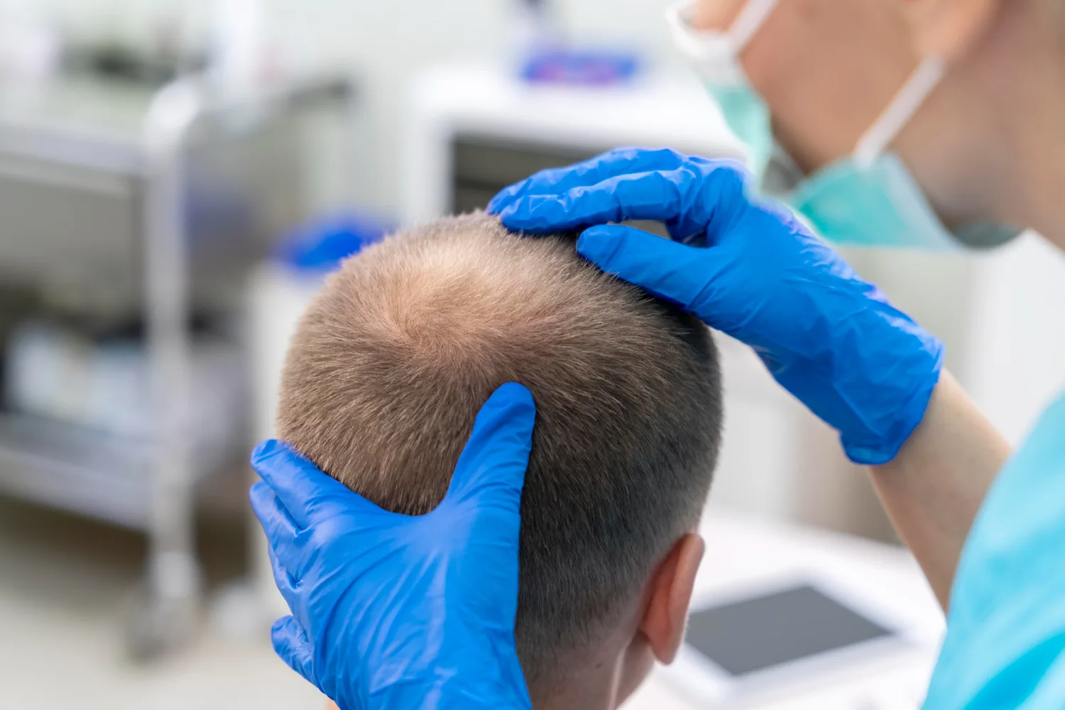
Donor Area Management During Hair Transplant
Why Is Donor Area Management Important During Hair Transplant?
Donor area management determines the long-term success of every hair transplant. Surgeons must extract follicles from a limited region while preserving density for the future. The donor area supplies all grafts for transplantation. Once surgeons remove a follicular unit, that exact unit never returns. This reality makes donor management the most critical factor in sustainable hair restoration.
The donor area sits at the back and sides of the scalp. These regions contain hair follicles that resist androgenetic miniaturization better than frontal or crown hair. Surgeons call this phenomenon donor dominance. Dr. Norman Orentreich first described donor dominance in 1959. His research showed that transplanted follicles keep their original characteristics even in new locations (Orentreich 1959). This principle allows surgeons to move permanent hair to balding areas.
However, donor dominance does not mean the donor area contains infinite grafts. Every patient owns a finite number of donor follicles. Poor extraction planning exhausts this supply. Overharvesting creates visible thinning, scarring, and patchy density. Patients then lose options for future procedures. Smart donor management balances immediate cosmetic improvement with long-term preservation. Surgeons must protect the donor reserve for potential future hair loss.
What Is the Donor Area in Hair Transplantation?
Where Is the Donor Area Located?
The donor area occupies the occipital and parietal regions of the scalp. These areas cover the back and sides of the head. Surgeons identify a specific region within this area called the safe donor zone. Dr. Walter Unger defined the safe donor zone through decades of clinical observation. His research established that this zone contains the most stable hair follicles throughout a patient's lifetime (Unger 1994).
The exact boundaries of the safe donor zone vary between individuals. Genetics control hair-loss patterns. Some men develop extensive baldness that creeps into the lower occipital region. Others maintain dense hair across the entire back of the scalp. Age also changes donor stability. Younger patients may show full donor density now but lose some of it later. Miniaturization can quietly affect donor follicles even before patients notice visible thinning.
Why Is the Donor Area Usually Chosen for Hair Transplantation?
Surgeons select the donor area for one primary reason. The follicles here resist androgenetic miniaturization better than follicles on the top of the head. Androgens shrink genetically vulnerable follicles over time. Donor follicles carry different genetic programming. They maintain their diameter and growth cycle even when transplanted to balding areas.
Donor hair also offers excellent density and favorable follicular-unit composition. Many donor follicles contain two or three hairs per unit. This characteristic maximizes visual coverage. Hair caliber matters too. Thick donor hair creates more visual density than fine hair. Surgeons evaluate all these factors before planning extraction.
Is the Donor Area Truly Permanent?
The term "permanent donor area" creates dangerous misconceptions. No donor region carries an absolute lifetime guarantee. Some follicles within the safe donor zone can miniaturize over time. This risk increases in patients with advanced androgenetic alopecia. Future hair-loss progression may slowly affect even relatively stable donor regions.
Surgeons must distinguish between relatively stable donor hair and truly permanent hair. Most donor follicles survive for decades. However, a small percentage may thin over time. This reality makes conservative planning essential. Surgeons cannot assume every donor follicle will last forever.
How Is the Donor Area Evaluated Before Hair Transplant Surgery?
Which Characteristics Determine Donor Hair Quality?
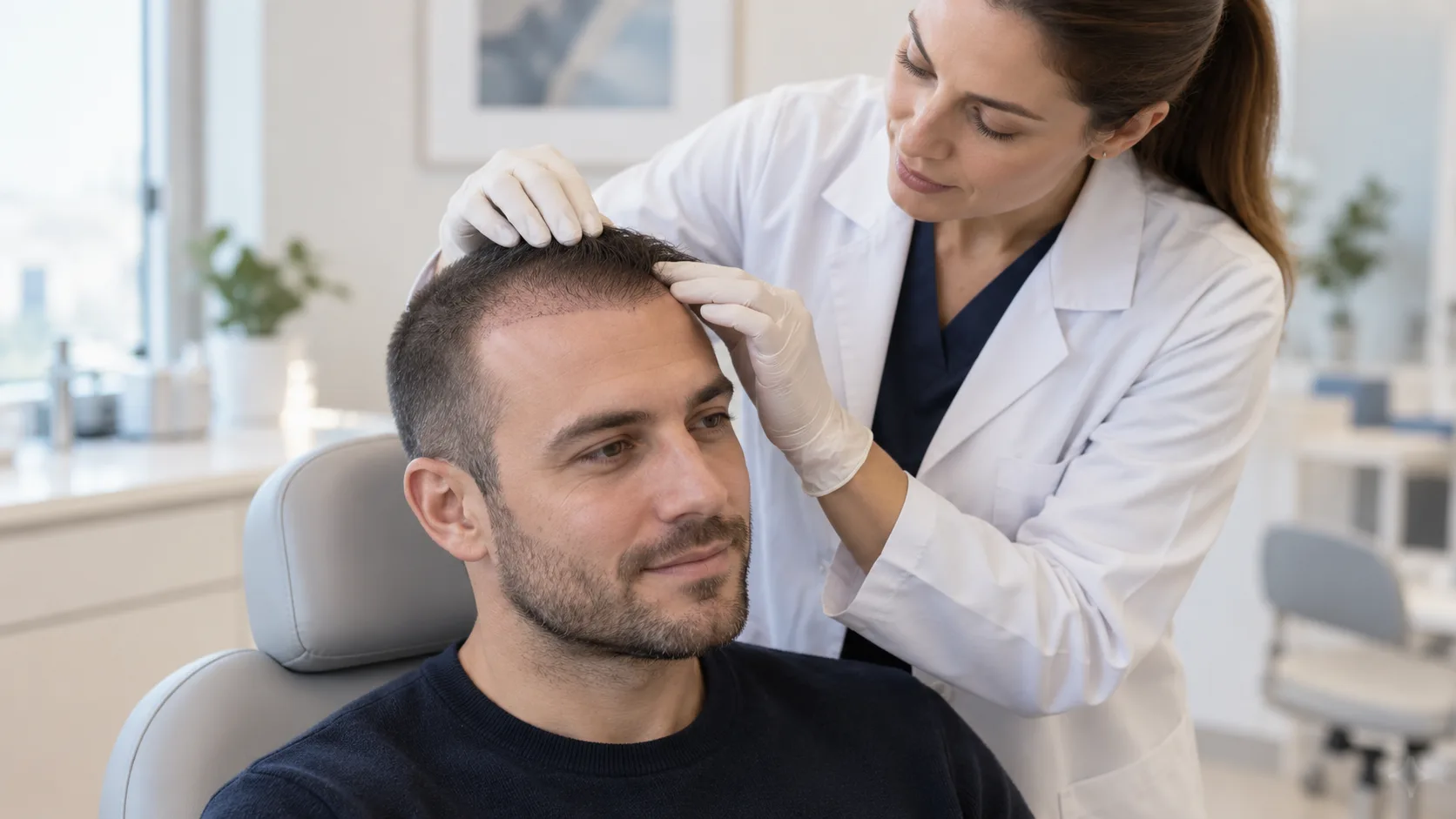
Surgeons evaluate multiple characteristics before harvesting any grafts. Each factor influences the final aesthetic result.
Hair density measures how many hairs grow per square centimeter. Higher density allows more graft extraction while maintaining visual fullness.
Hair caliber refers to the thickness of individual hair shafts. Thick hair creates more visual coverage than thin hair.
Follicular-unit composition describes how many hairs exist within each follicular unit. Units with two or three hairs provide better coverage than single-hair units.
Hair-to-scalp contrast affects how noticeable thinning appears. Dark hair on light skin shows more contrast than light hair on light skin.
Distribution of follicular units reveals whether density remains uniform or varies across the donor region.
Miniaturization indicates whether some donor follicles have already started shrinking.
Scalp condition includes elasticity, scarring from previous procedures, and overall skin health.
Donor-zone dimensions determine the total surface area available for extraction.
How Does Trichoscopy Help Assess the Donor Area?
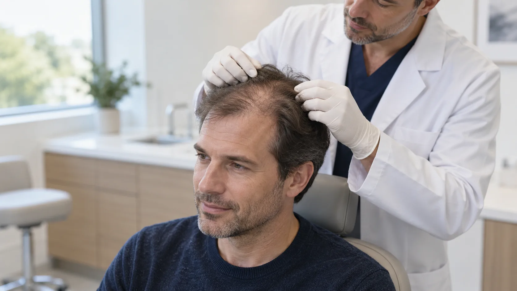
Trichoscopy provides a magnified view of the scalp. This tool helps surgeons identify problems invisible to the naked eye. Trichoscopy reveals miniaturized follicles that signal early donor instability. It measures exact follicular density across different donor regions. Surgeons can assess hair shaft caliber with precision. The tool also detects heterogeneous donor density, where some areas contain more hair than others. Trichoscopy maps potentially unstable donor regions before extraction begins. Dr. Antonella Tosti advanced trichoscopy applications in hair disorders. Her research demonstrated its value in preoperative donor evaluation (Tosti 2007).
How Does Family History Affect Donor Area Planning?
Family history predicts future hair-loss patterns. Surgeons examine the patient's father, grandfathers, and maternal relatives. Advanced baldness in close relatives suggests aggressive future progression. This information helps surgeons plan conservatively.
Younger patients require especially careful evaluation. Their future pattern of hair loss remains uncertain. A 25-year-old may show only early recession now but develop extensive baldness by age 50. Surgeons must preserve donor reserves for these future needs. Current donor appearance does not guarantee future donor stability.
Can Medical Hair-Loss Treatment Influence Donor Area Planning?
Medical treatments like finasteride and minoxidil can stabilize miniaturizing native hair. Finasteride blocks dihydrotestosterone conversion. Minoxidil improves follicular blood flow. These medications may slow hair loss and preserve existing density.
Treatment response influences surgical timing. A patient responding well to medication might delay surgery or require fewer grafts. However, medical therapy does not eliminate biological uncertainty. Medications cannot guarantee permanent follicular stability. Surgeons must still plan conservatively even when patients use these treatments. Dr. Keith D. Kaufman published pivotal research on finasteride efficacy in men with androgenetic alopecia (Kaufman 1998).
How Many Grafts Can Safely Be Harvested From the Donor Area?
Why Is Donor Hair Considered a Finite Resource?
Extracted follicular units never regenerate in their original donor sites. This fact defines donor hair as a finite, non-renewable resource. When a surgeon removes a follicular unit, that specific unit disappears forever from the donor region. Surrounding follicles continue growing. However, the extracted unit itself does not return.
Patients must understand this biological reality. Every graft represents part of a lifetime donor reserve. Surgeons should treat each follicle as a precious resource. Wasteful extraction depletes this reserve permanently.
Which Factors Determine Safe Donor Graft Numbers?
Multiple factors control how many grafts surgeons can safely harvest.
Baseline donor density sets the upper limit. A patient with 80 follicular units per square centimeter offers more grafts than one with 50.
Hair caliber affects visual impact. Thick hair requires fewer grafts for the same visual result.
Follicular-unit density measures how many units exist per square centimeter.
Scalp surface area determines the total donor region size. Larger donor zones contain more total grafts.
Degree and pattern of hair loss influence how many grafts the patient needs now and in the future.
Patient age matters because younger patients need more reserves for future procedures.
Expected future progression requires surgeons to save grafts for later hair loss.
Previous transplantation procedures reduce available donor supply.
Extraction technique affects how efficiently surgeons harvest without damaging surrounding follicles.
Distribution of extraction sites determines whether harvesting creates visible thinning.
Why Should Graft Count Alone Not Determine Surgical Planning?
Graft quantity differs from cosmetic coverage. A patient with 2,000 thick, multi-hair grafts achieves better coverage than a patient with 3,000 fine, single-hair grafts. Coverage value depends on hair caliber, angulation, and distribution.
Maximizing graft numbers does not automatically maximize aesthetic results. Some clinics aggressively harvest excessive grafts to advertise high numbers. This approach depletes donor reserves and creates visible thinning. Smart planning prioritizes cosmetic coverage while preserving donor density for the future.
How Does Donor Area Management Differ Between FUE and FUT?
How Does FUE Affect the Donor Area?
Follicular Unit Extraction (FUE) removes individual follicular units one by one. Surgeons use small circular punches to extract each unit. This technique creates tiny circular wounds across the donor region.
Punch size matters enormously. Smaller punches (0.8-1.0 mm) create less visible scarring than larger punches. Extraction depth must match follicle angulation. Surgeons must follow the natural tilt of each hair to avoid transection. Proper spacing between extraction sites preserves surrounding follicles and maintains visual density.
Poor FUE technique causes diffuse donor thinning. Excessive extraction from a limited region creates a "moth-eaten" appearance. Even small scars accumulate when surgeons harvest too aggressively.
How Does FUT Affect the Donor Area?
Follicular Unit Transplantation (FUT) removes a strip of hair-bearing scalp from the donor region. Surgeons then dissect this strip into individual follicular units under microscopes. The technique leaves a linear donor scar.
Surgical closure technique determines scar quality. Skilled surgeons use trichophytic closure to minimize visible scarring. This method allows hair to grow through the scar line. FUT preserves surrounding donor follicles because extraction occurs from a single strip rather than scattered across the entire donor region.
However, FUT limits future strip procedures. Each strip removes a section of scalp. Patients cannot undergo unlimited FUT procedures. The linear scar also restricts how short patients can wear their hair.
Which Technique Is Better for Preserving the Donor Area?
Neither FUE nor FUT offers universal superiority for donor preservation. The better technique depends on patient characteristics, surgical planning, harvesting strategy, and surgeon expertise.
FUE creates diffuse extraction effects across the donor region. FUT creates a single linear scar. Both techniques draw from the same finite biological donor supply. A skilled FUE surgeon preserves donor density better than an unskilled FUT surgeon, and vice versa. Patients should select an experienced surgeon rather than a specific technique.
Factor | FUE Donor Impact | FUT Donor Impact |
Scar pattern | Multiple tiny dots | Single linear scar |
Hair length restriction | Minimal if well-executed | May require longer hair to conceal |
Surrounding follicle preservation | Risk of adjacent damage | Preserves follicles outside strip |
Future strip procedures | Possible | Limited by scar tissue |
Healing time | 7-14 days for surface | 10-14 days for suture line |
Donor thinning risk | Diffuse if overharvested | Localized to strip area |
What Is Overharvesting of the Donor Area?
What Causes Donor Area Overharvesting?
Overharvesting occurs when surgeons extract too many grafts from the donor region. Several specific mistakes cause this problem.
Excessive graft extraction beyond safe limits depletes donor density. Extraction sites placed too close together damage surrounding follicles and create visible gaps. Excessive harvesting from a limited region concentrates damage in one area. Large graft sessions without adequate donor assessment ignore individual biological limitations. Repeated procedures without long-term planning exhaust reserves prematurely.
What Does an Overharvested Donor Area Look Like?
An overharvested donor area shows clear visual signs. Patchy density appears where extraction removed too many follicles. Visible gaps separate remaining hair clumps. Diffuse thinning reduces overall donor density. Some patients develop a "moth-eaten" appearance with scattered bald spots. Increased scalp visibility occurs because remaining hair cannot cover the skin adequately. Scarring becomes visible, especially with short hair. Patients lose the ability to wear their hair very short.
Can Overharvesting Become Visible Years After Surgery?
Overharvesting can appear immediately or develop gradually. Some patients notice donor thinning within months. Others see problems emerge years later.
Cumulative tissue trauma changes the donor environment over time. Extraction damages surrounding follicles even when surgeons avoid direct injury. Blood supply changes and scar tissue formation affect nearby follicles. A donor area may look acceptable during early recovery but thin progressively as surrounding follicles weaken.
Long-term follow-up matters more than early recovery appearance. Surgeons and patients must evaluate donor density at six months, one year, and beyond.
How Can Surgeons Prevent Donor Area Overharvesting?
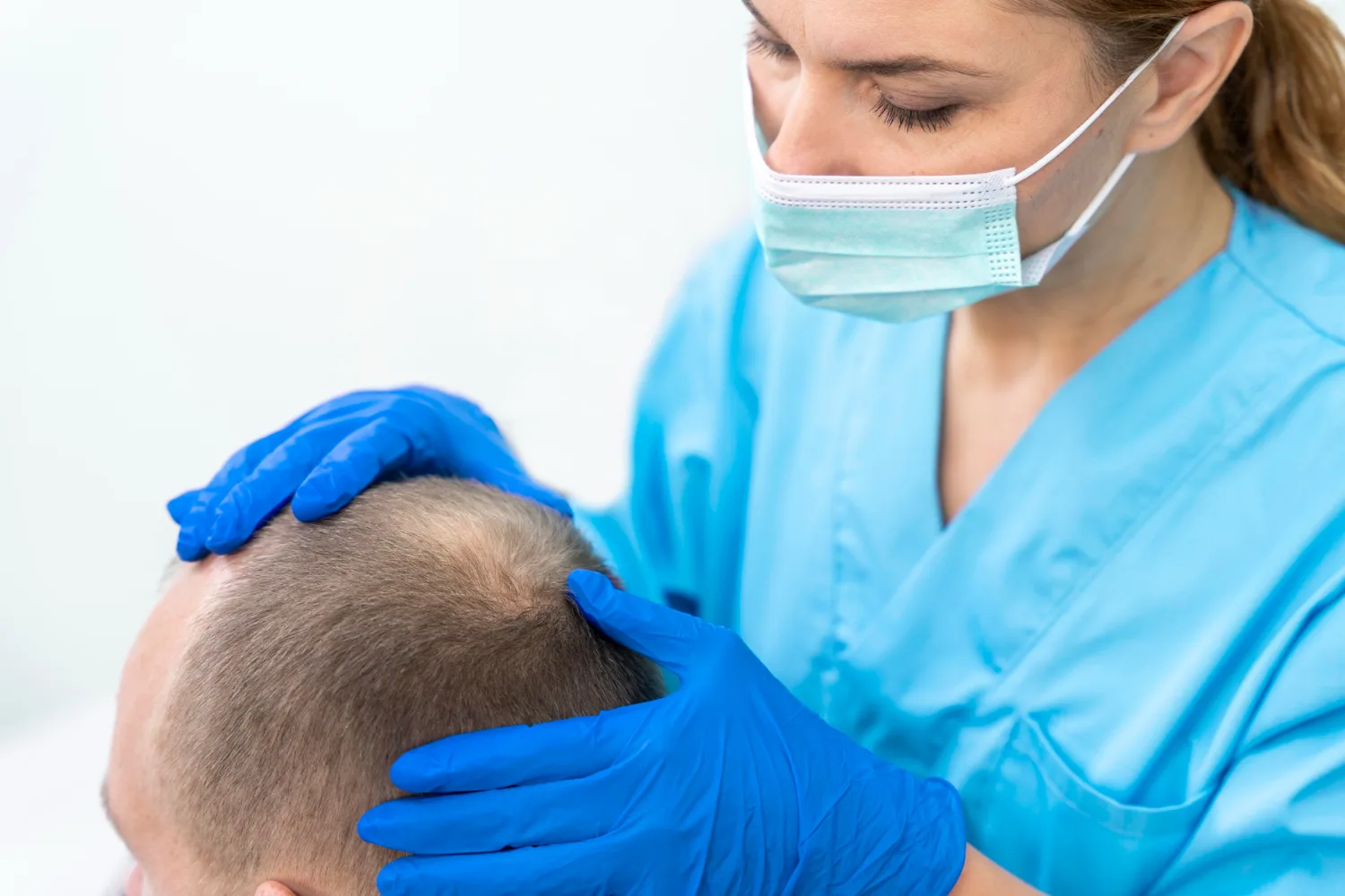
How Should Extraction Sites Be Distributed?
Proper distribution prevents visible donor depletion. Surgeons must maintain appropriate spacing between extraction sites. This spacing protects adjacent follicles from trauma. Surgeons should avoid excessive concentration in any single region. They must preserve overall visual donor density even when harvesting large numbers. Future procedures require careful consideration during initial extraction pattern design. A well-distributed pattern leaves uniform density across the entire safe donor zone.
Why Is Conservative Graft Harvesting Important?
Conservative harvesting protects remaining follicular units. It reduces visible donor depletion and maintains future surgical options. It also protects the natural appearance of the donor region.
Surgeons should resist pressure to maximize graft numbers. A session of 1,500 well-placed grafts often creates better results than 3,000 poorly distributed grafts. Conservative extraction preserves the donor reserve for future needs.
Why Should Large Graft Sessions Be Planned Carefully?
Biological limits govern extensive harvesting. Human scalps contain finite follicular units. Surgeons cannot safely extract unlimited grafts in one session.
The relationship between graft numbers and donor reserve demands careful calculation. "More grafts" does not automatically mean better outcomes. Some patients benefit from staged treatment. Two moderate sessions often produce better results than one massive session that damages the donor region.
What Happens to the Donor Area Immediately After Hair Transplantation?
What Are Normal Donor Area Symptoms?
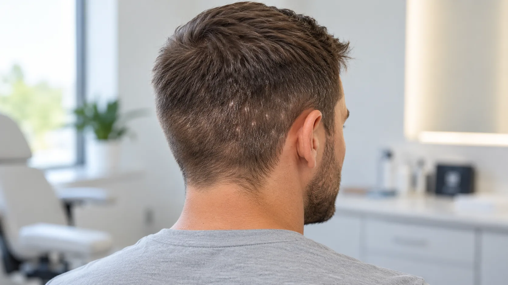
Patients should expect several normal symptoms after donor harvesting. Redness appears across the extraction or incision sites. Mild soreness or sensitivity persists for several days. Itching commonly occurs as healing progresses. Small scabs form at each extraction site. Temporary swelling may affect the donor region and surrounding areas. Temporary changes in hair density occur because of postoperative inflammation and short hair length.
Why Does the Donor Area Form Small Scabs?
Scabs form as part of the natural healing response. FUE creates micro-wounds when punches penetrate the skin. Each extraction site bleeds slightly and then clots. These clots dry into small scabs. The scabs protect underlying tissue during early healing. They naturally separate and shed as the skin regenerates beneath them. Patients should not force this process.
How Long Does Donor Area Healing Take?
Healing progresses through predictable stages.
Days 1-3 bring redness, tenderness, and early scabbing. Patients should follow their surgeon's cleaning instructions during this period.
Days 4-7 show progressive scab shedding. Most surface scabs detach naturally during this window.
Days 8-14 bring decreasing sensitivity and normalization of skin appearance. The donor region starts looking more natural.
After approximately two weeks, visible healing advances substantially. However, deeper tissue remodeling continues for months beneath the surface.
Recovery Phase | Timeline | Expected Appearance |
Acute healing | Days 1-3 | Redness, tenderness, scabbing |
Early recovery | Days 4-7 | Scabs shedding, decreasing redness |
Surface healing | Days 8-14 | Normalizing skin, reduced sensitivity |
Deep remodeling | Weeks 2-12 | Surface looks healed, tissue still remodeling |
Does Donor Hair Grow Back After a Hair Transplant?
Do Extracted Follicles Regrow in the Donor Area?
Extracted follicular units never regrow in their original donor locations. This fact confuses many patients. When a surgeon removes a follicle, that specific follicle disappears permanently from the donor site. The extracted graft then grows after transplantation in the recipient area.
Some patients mistake continued growth of surrounding follicles for regrowth of extracted ones. Neighboring follicles that survived extraction continue their normal growth cycle. This creates the impression that hair "grew back." True follicular regeneration does not occur in the donor area after standard extraction.
Why Can the Donor Area Look Fuller Again After Surgery?
Several factors create the appearance of fuller donor hair. Surrounding native follicles continue growing normally. Postoperative redness fades, reducing the contrast between hair and skin. Scabs disappear, revealing the natural scalp surface. Hair length increases as trimmed donor hair grows out. Remaining follicles gradually blend together, creating more uniform visual density.
How Should the Donor Area Be Managed During Recovery?
How Should the Donor Area Be Washed?
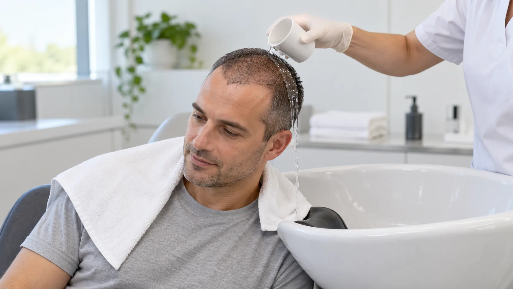
Patients must follow their surgeon's individualized postoperative instructions. Each surgeon designs washing protocols based on technique and patient factors. Generally, patients should use gentle cleansing techniques. They must avoid aggressive rubbing or scratching during early healing. Mild shampoo applied with fingertips usually suffices. Patting the area dry with a soft towel prevents unnecessary friction.
When Can the Donor Area Be Touched?
Gentle contact during washing differs from scratching or rubbing. Patients can touch the donor area lightly for cleaning purposes. However, they must avoid picking, scratching, or rubbing the healing tissue. Protection of healing tissue remains essential for the first two weeks.
Should Patients Pick Donor Area Scabs?
Patients should never pick donor area scabs. Scabs detach naturally when underlying skin heals sufficiently. Premature picking tears healing tissue and damages follicles. It also introduces bacteria and risks infection. Picking can cause unnecessary trauma that leads to visible scarring or patchy density.
When Can Patients Resume Normal Hair Care?
Timing depends on surgical technique and individual healing. Most patients gradually return to normal washing within two weeks. Styling and hair-care practices resume under clinical guidance. Surgeons typically clear patients for normal routines after surface healing completes. Patients should confirm timing with their surgeon rather than making assumptions.
What Donor Area Complications Should Patients Watch For?
Which Donor Area Changes Are Usually Temporary?
Several symptoms cause concern but resolve naturally. Mild redness fades within days to weeks. Scabbing disappears as healing progresses. Tenderness diminishes progressively. Itching signals healing but should not trigger scratching. Temporary visual thinning occurs because of short hair length and inflammation.
What Symptoms May Require Medical Evaluation?
Some symptoms indicate problems that need professional attention. Increasing pain rather than improving pain suggests complications. Persistent or worsening redness beyond two weeks requires evaluation. Significant swelling that increases rather than decreases needs assessment. Pus or drainage signals possible infection. Severe folliculitis with multiple painful bumps requires treatment. Excessive bleeding after the first day needs immediate attention. Delayed wound healing beyond normal timelines concerns surgeons. Progressive donor-area thinning unrelated to normal healing suggests overharvesting.
What Is Donor Area Folliculitis?
Postoperative folliculitis means inflammation of hair follicles in the donor region. It appears as small red bumps or pustules around hair follicles. Mild cases resolve with proper cleaning. Severe or widespread folliculitis requires medical treatment. Patients must distinguish expected postoperative irritation from signs of infection. Expected irritation remains mild and improves daily. Infection shows increasing severity, spreading redness, or pus formation.
Symptom | Usually Normal | Needs Evaluation |
Mild redness | First 1-2 weeks | Persisting beyond 3 weeks |
Small scabs | First 7-10 days | Not improving by day 14 |
Mild tenderness | First 5-7 days | Increasing after day 3 |
Itching | First 2 weeks | Severe or with rash |
Slight swelling | First 3-5 days | Increasing after day 5 |
What Is the Difference Between Temporary Donor Thinning and Permanent Donor Depletion?
Why Can the Donor Area Look Thin Immediately After Surgery?
Temporary thinning has several causes. Short hair length following shaving makes density appear lower. Postoperative inflammation causes redness that increases scalp visibility. Scabbing creates texture changes that affect visual appearance. Temporary changes in hair visibility resolve as healing completes and hair grows longer.
What Does Permanent Donor Depletion Look Like?
Permanent depletion shows distinct characteristics. Persistent gaps remain visible months after surgery. Reduced follicular density becomes measurable with trichoscopy. Patchy extraction patterns reveal where surgeons concentrated harvesting. Visible scarring appears, especially with short hair. Progressive thinning continues unrelated to the normal healing process.
When Should Donor Density Be Reassessed?
Surgeons should assess donor density at multiple intervals. Early postoperative evaluation occurs at one to two weeks. Intermediate follow-up happens at six to twelve months. Long-term evaluation continues as native hair loss progresses. Annual assessments help detect gradual donor changes.
Can Donor Area Damage Be Repaired?
Can an Overharvested Donor Area Return to Its Original Density?
Biological limitations prevent full reversal of donor depletion. Extracted follicles never return. Substantial donor depletion cannot completely reverse. Surgeons can improve appearance through cosmetic camouflage. However, true follicular restoration remains impossible. Patients must understand this limitation before seeking repair.
Can Hair Be Transplanted Into an Overharvested Donor Area?
Surgeons can perform donor-area repair in selected cases. They transplant grafts into visibly depleted regions. However, the remaining donor supply limits this approach. Surgeons cannot create new follicles. They can only redistribute existing ones.
Some patients use alternative donor sources. Beard hair offers additional grafts when scalp donor hair runs low. Body hair provides another option in carefully selected patients. These alternatives carry significant limitations.
What Are the Limitations of Beard or Body Hair for Donor Repair?
Beard and body hair differ substantially from scalp hair. Hair caliber varies, often creating texture mismatches. Growth cycles differ, with body hair spending more time in the resting phase. Shaft characteristics vary in curl, color, and diameter. Aesthetic integration requires careful planning. Alternative donor hair cannot fully replace scalp donor hair in most cases.
Can Beard and Body Hair Be Used When Scalp Donor Hair Is Limited?
When Is Beard Hair Considered as an Alternative Donor Source?
Beard hair helps when scalp donor supply proves insufficient. Selected repair procedures benefit from beard grafts. Surgeons must match hair characteristics to the recipient area. Beard hair typically grows thicker and curlier than scalp hair. It works best for adding density rather than creating hairlines.
What Are the Limitations of Body Hair?
Body hair presents several challenges. Different growth cycles mean body hair sheds and regrows unpredictably. Variable shaft characteristics create texture inconsistencies. Differences in length and behavior make blending difficult. Cosmetic integration remains less predictable than with scalp hair. Dr. Raymond Woods pioneered body hair transplantation techniques. His research documented both possibilities and limitations of this approach (Woods 2002).
How Does Long-Term Hair Loss Affect Donor Area Management?
Why Should Future Hair Loss Be Considered Before the First Transplant?
Hair transplantation does not stop underlying androgenetic alopecia. Native hair continues miniaturizing even after surgery. A patient may transplant hair into the frontal region while crown hair continues thinning. Future surgical requirements will arise.
Donor planning must account for these future needs. Surgeons should reserve grafts for subsequent procedures. A patient who uses all donor grafts for the first surgery loses options when new bald areas develop.
Why Is Long-Term Planning Especially Important in Younger Patients?
Younger patients face unique challenges. Their future pattern of hair loss remains uncertain. A 22-year-old may develop only mild recession or progress to extensive baldness. Greater lifetime need for donor resources demands conservative planning. Young patients risk exhausting donor reserves prematurely if surgeons treat only immediate concerns.
Dr. Robert Bernstein emphasized this principle in his research on hair transplant planning. He advocated for conservative approaches in young patients to preserve options for future hair loss (Bernstein 2005).
How Can Medical Management Complement Surgical Donor Management?
Medical therapy maintains native hair and reduces future surgical needs. Finasteride and minoxidil slow miniaturization. Dutasteride offers stronger DHT suppression for some patients. Low-level laser therapy and platelet-rich plasma provide additional support.
Stabilization supports long-term surgical planning. A patient who maintains existing hair needs fewer grafts over time. However, medication cannot guarantee permanent follicular stability. Surgeons must still plan conservatively even with medical treatment.
What Does Good Donor Area Management Look Like Over the Long Term?
Excellent donor management follows clear principles. Accurate preoperative donor assessment establishes baseline density and quality. Conservative extraction preserves remaining follicles. Appropriate distribution of grafts prevents visible thinning. Preservation of donor density maintains natural appearance. Monitoring of native hair loss detects progression early. Appropriate postoperative care supports healing. Consideration of future transplantation needs guides every decision. Long-term documentation with standardized photographs and trichoscopic measurements tracks changes objectively.
Frequently Asked Questions About Donor Area Management During Hair Transplant
Does the donor area grow back after a hair transplant?
No. Extracted follicles never regrow in the donor area. Surrounding follicles continue growing, but removed units disappear permanently.
How long does the donor area take to heal?
Surface healing completes within two weeks. Scabs shed by day 7-10. Redness fades by week 2-3. Deep tissue remodeling continues for months.
Is redness in the donor area normal after hair transplantation?
Yes. Mild redness remains normal for the first two weeks. Persistent or worsening redness beyond this period requires medical evaluation.
Can the donor area become permanently thin?
Yes. Overharvesting causes permanent donor depletion. Excessive extraction damages density irreversibly.
What causes an overharvested donor area?
Excessive graft extraction, poor site distribution, large sessions without proper assessment, and repeated procedures without long-term planning cause overharvesting.
How many grafts can be safely removed from the donor area?
Safe numbers vary by individual. Factors include donor density, hair caliber, scalp size, and future loss expectations. Surgeons should prioritize quality over quantity.
Can an overharvested donor area be repaired?
Surgeons can improve appearance through camouflage techniques and limited grafting. However, full restoration of original density remains impossible.
Is FUE safer for the donor area than FUT?
Neither technique offers universal superiority. Both affect the donor region differently. Surgeon skill matters more than technique choice.
Can beard hair be used if scalp donor hair is insufficient?
Yes. Beard hair serves as an alternative donor source in selected cases. However, texture and growth differences limit its applications.
Can the donor area continue to thin after a hair transplant?
Yes. Some donor follicles may miniaturize over time. Future hair-loss progression can affect even relatively stable donor regions.
How can patients protect their donor area after surgery?
Patients should follow surgeon instructions, wash gently, avoid picking scabs, protect from sun exposure, and attend all follow-up appointments.
Conclusion: Why Is Donor Area Management Essential for a Sustainable Hair Transplant?
Donor area management determines whether hair transplantation succeeds over a lifetime. Donor hair remains limited. Extracted follicles never regenerate in their original sites. This biological reality demands respect and careful planning.
Successful donor management requires multiple components. Accurate preoperative assessment identifies available resources. Conservative harvesting preserves density for the future. Appropriate extraction distribution maintains natural appearance. Postoperative care supports proper healing. Long-term planning accounts for progressive hair loss.
The objective extends beyond maximizing transplanted grafts. Smart surgeons optimize cosmetic coverage while preserving donor reserves. They balance immediate improvement against future needs. Patients benefit most when surgeons treat donor hair as the precious, finite resource that it truly is.
References
Bernstein, Robert M., and William R. Rassman. "Follicular Unit Transplantation: 2005." Dermatologic Clinics, vol. 23, no. 3, 2005, pp. 393-414.
Kaufman, Keith D., et al. "Finasteride in the Treatment of Men with Androgenetic Alopecia." Journal of the American Academy of Dermatology, vol. 39, no. 4, 1998, pp. 578-589.
Orentreich, Norman. "Autografts in Alopecias and Other Selected Dermatological Conditions." Annals of the New York Academy of Sciences, vol. 83, no. 3, 1959, pp. 463-479.
Tosti, Antonella, et al. "Trichoscopy: A New Tool for Diagnosing Hair Loss." Dermatologic Clinics, vol. 25, no. 3, 2007, pp. 357-364.
Unger, Walter P. "The Current Status of Hair Transplantation." Dermatologic Clinics, vol. 12, no. 4, 1994, pp. 679-686.
Woods, Raymond. "Body Hair Transplantation: A New and Emerging Frontier." Hair Transplant Forum International, vol. 12, no. 4, 2002, pp. 121-124.