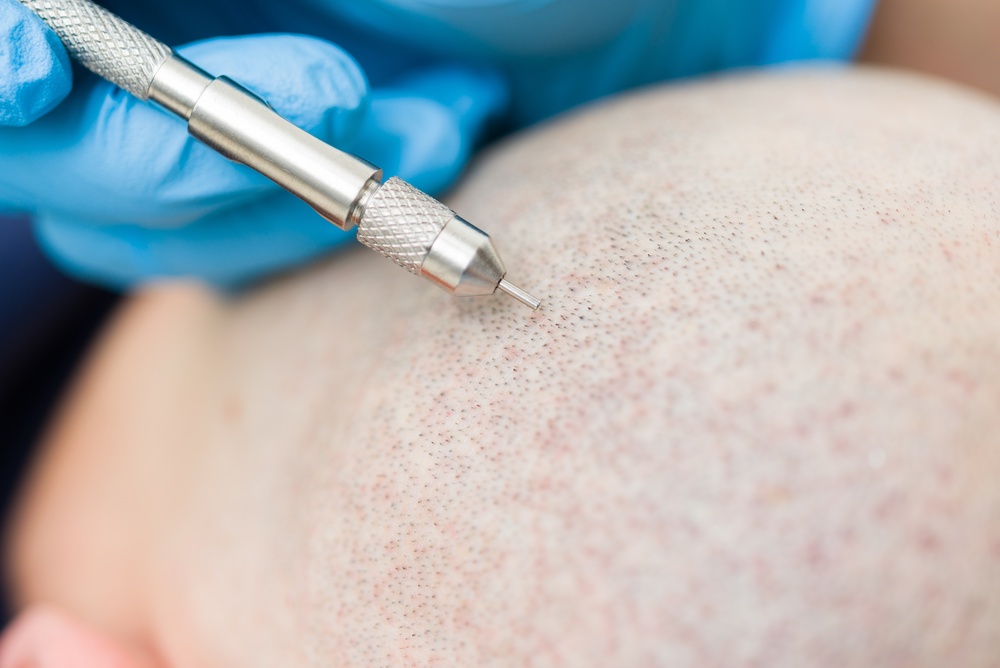
Canal opening creates tiny recipient sites in the scalp. Surgeons place extracted grafts into these sites. This single step determines growth direction, density, and final appearance.
Follicular Unit Extraction, or FUE, stands as the leading minimally invasive method for hair restoration today. The procedure breaks into three clear stages. First, the surgeon extracts individual follicular units from the donor areaThe Source of Restoration The donor area plays a critical role in hair transplantation, as it serves as the source.... Second, the surgeon opens canals in the recipient zone. Third, the surgeon implants the grafts into those canals. Many patients focus only on extraction, but canal opening actually controls every visual outcome. The angle, depth, and spacing of these micro-incisions dictate whether the new hair looks natural or artificial. Bernstein and Rassman (2005) established that recipient site creation directly impacts graft survival rates and aesthetic results. Without precise canal opening, even perfect grafts fail to produce satisfactory coverage.
What Does Canal Opening Mean in Modern Hair Restoration?
Canal opening means making micro-incisions in the bald area. These incisions receive the extracted follicular units. Each canal serves as a tiny pocket for one graft.
Surgeons call this step recipient site creation. They use blades or needles to cut small slits or holes in the scalp. These openings must match the exact size of the graft. If the canal is too wide, the graft moves. If the canal is too narrow, the graft crushes. The surgeon must also align each canal with the natural hair flow. This alignment ensures the new hair lies flat and follows the same pattern as the surrounding native hair. Uebel (1997) demonstrated that recipient sites must respect the anatomical distribution of follicular units to achieve natural density. Canal opening is not random cutting. It is a calculated architectural process that rebuilds the hairline and fills bald zones with mathematical precision.
How Does Canal Opening Support Graft Survival?
Proper canal opening protects blood supply. It also ensures correct graft placement and orientation. These two factors keep follicles alive after transplantation.
Each graft contains living tissue. It needs oxygen and nutrients from the scalp. When a surgeon creates a clean, well-sized canal, the surrounding blood vessels stay intact. The graft then taps into this vascular network quickly. A rough or oversized canal damages nearby capillaries. This damage starves the graft. Rose (2013) confirmed that careful recipient site preparation preserves scalp vascularization and improves follicle integration. The canal also holds the graft at the right depth. If the graft sits too deep, it buries. If it sits too shallow, it pops out. Correct depth anchors the graft and lets it feed from the tissue below. Canal opening, therefore, acts as the bridge between extraction and growth.
Why Does Natural Hair Direction Matter During Canal Creation?
Natural hair direction controls angle and tilt. Surgeons must copy this pattern for realistic results. Ignoring natural flow produces hair that sticks out or lies unnaturally.
Human hair does not grow straight up. It emerges from the scalp at specific angles. Hair at the crown swirls. Hair at the temples points backward. Hair at the hairline points forward and downward. The surgeon studies these patterns before picking up any blade. Each canal must mirror the angle of the native hair in that exact spot. If the surgeon sets the canal at the wrong tilt, the new hair grows in a different direction. This mistake creates a pluggy or unnatural look. Gho et al. (2011) emphasized that mimicking natural follicular unit distribution remains essential for seamless integration. The canal angle must also change as the surgeon moves across the scalp. One fixed angle does not work for all zones.
What Technical Principles Guide Canal Opening?
Canal opening follows four core rules. Surgeons control angle, depth, density, and native hair protection. These rules work together to create a natural result.
What Angle Should Surgeons Use During Canal Opening?
Surgeons use 30 to 45 degrees in most areas. The angle varies by scalp region. This range matches natural hair emergence.
The frontal hairline demands a low angle. New hairs here should lie flat against the forehead. A steep angle makes the hair stick up like bristles. The mid-scalp and crown need slightly steeper angles. The swirl at the crown requires radial placement. The surgeon adjusts the blade tilt constantly. Cole (2013) noted that maintaining consistent angle across hundreds or thousands of canals tests surgical skill. Each canal must match its neighbors. Even small deviations create visible irregularities. The blade enters the skin at the set angle and exits at the same tilt. This consistency creates a uniform carpet of hair.
How Deep Should Canals Go?
Canals must match graft length. Typical depth ranges from 4 to 6 millimeters. This depth anchors the graft without burying it.
A graft contains the follicle bulb and surrounding tissue. The bulb needs coverage. The surface needs exposure. The canal must reach deep enough to hold the graft firmly. If the surgeon cuts too shallow, the graft protrudes. It dries out and dies. If the surgeon cuts too deep, the graft sinks below the surface. It fails to receive adequate oxygen. The surgeon measures grafts under a microscope before opening canals. He then selects a blade or needle that matches. Avram et al. (2017) found that depth mismatch causes graft popping and poor growth rates. Precise depth control separates expert results from average outcomes.
How Do Surgeons Plan Canal Density?
Density planning balances coverage with blood supply. Surgeons avoid overpacking grafts. Too many grafts in one area kills tissue.
Density means how many grafts fit into one square centimeter. The scalp can only support a certain number. If the surgeon places grafts too close, the blood vessels cannot feed them all. The tissue turns pale. Some grafts die. This complication causes patchy growth. Most surgeons place 30 to 40 grafts per square centimeter in a single session. Some push to 50 in select cases. Haber et al. (2015) warned that excessive density risks recipient site necrosis and compromises graft survival. The surgeon must also plan density based on the donor supply. A patient with limited grafts needs strategic placement. The surgeon maps high-density zones and transition zones before making the first cut.
Why Must Surgeons Protect Native Hair?
Existing hair follicles sit near new sites. Sharp blades can cut these native roots. Surgeons must avoid damaging surviving hair.
Many patients undergo FUE to thicken thinning areas. They still have native hair in these zones. The surgeon must place new canals between existing follicles. A careless incision transects a native root. This kills a hair that was already growing. The patient loses more hair than he gains. The surgeon uses magnification to see native follicles. He steers blades around them. Unger and Shapiro (2011) stressed that preserving native hair maintains overall density and prevents iatrogenic hair loss. This protection requires patience and visual precision. The surgeon cannot rush canal opening in areas with remaining hair.
What Instruments Do Surgeons Use for Canal Opening?
Surgeons choose from four main tools. Each tool creates canals differently. The choice affects precision, healing, and density.
|
Instrument |
Incision Size |
Healing Speed |
Density Potential |
Cost |
|
Sapphire BladeA Modern Innovation in Hair Transplants The sapphire blade is an advanced tool used to create precise incisions during hair... |
Very Small |
Fast |
High |
Higher |
|
Steel Blade |
Medium |
Standard |
Moderate |
Lower |
|
Micro-Needle |
Ultra-Fine |
Fast |
High |
Moderate |
|
Implanter Pen |
Variable |
Fast |
High |
Higher |
Why Do Surgeons Prefer Sapphire Blades?
Sapphire blades make sharper, smaller cuts. These cuts heal faster and allow higher density. The blade material reduces tissue trauma.
Sapphire is a hard crystal. It holds an edge better than steel. Surgeons shape sapphire blades into V-tips or U-tips. These tips enter the scalp with minimal resistance. The incision stays narrow. A narrow incision means less bleeding. It also means the surgeon can place canals closer together. This closer spacing creates fuller hair. Doganay et al. (2019) reported that sapphire blade incisions produce less postoperative crusting and faster epithelial healing. Patients see less scabbing. The scalp returns to normal appearance sooner. The sharpness also reduces the force needed to penetrate the skin. Less force means less surrounding tissue damage. For these reasons, sapphire blades have become the premium choice in modern FUE.
How Does the Steel Blade Method Work?
Steel blades create traditional slit incisions. This method costs less but leaves slightly larger wounds. It remains common in many clinics.
A steel blade is a flat metal edge. The surgeon presses it into the scalp to make a linear slit. Steel blades come in different widths. The surgeon picks a width that matches the graft. However, steel dulls faster than sapphire. A dull blade tears rather than cuts. This tearing increases wound size. Larger wounds heal slower. They also create more visible scarring if the patient shaves his head. Despite these drawbacks, steel blades offer affordability. Many clinics achieve good results with sharp steel blades and skilled hands. The technique has served hair transplantationHair transplantation is a surgical procedure that involves the extraction of hair follicles from a designated donor site, followed by... for decades. It still works when the surgeon controls depth and angle carefully.
What Is the Micro-Needle Technique?
Micro-needles make ultra-fine channels. Surgeons customize diameter for each graft size. This technique suits patients with specific hair types.
A micro-needle is a hollow or solid tube with a tiny tip. Diameters range from 0.6 mm to 1.0 mm. The surgeon selects the needle based on the graft width. A single-hair graft gets a 0.6 mm channel. A three-hair graft gets a 0.9 mm channel. This customization prevents wiggling or compression. The needle creates a circular opening. Circular openings close evenly during healing. Micro-needles also allow rapid canal creation. The surgeon can make thousands of canals in a short time. However, the surgeon must control depth manually. Without a depth guard, the needle can go too deep. This tool demands steady hands and constant focus.
How Does the Implanter Pen Create Canals?
The implanter pen cuts and implants in one motion. This reduces graft handling. The technique is called Direct Hair Implantation, or DHI.
An implanter pen looks like a thick syringe. It holds a graft in its chamber. The surgeon presses the tip against the scalp. The tip pierces the skin and creates a canal. Then the pen releases the graft into that canal. The surgeon does not touch the graft with forceps. This lack of handling protects the fragile follicle. Bicer (2018) showed that DHI reduces graft trauma and improves survival in select patient populations. The pen also controls depth mechanically. A stopper prevents over-insertion. However, the pen works best with specific graft types. Very curly or bulky grafts may not fit smoothly. The surgeon must also work quickly. The graft sits outside the body while the surgeon positions each pen load.
What Happens During the FUE Procedure?
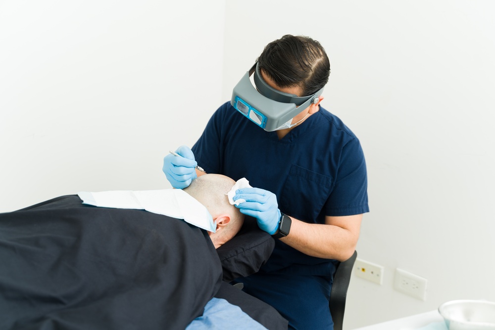
FUE follows a strict sequence. Each stage depends on the last. Canal opening sits at the center of this workflow.
How Do Surgeons Plan Before Canal Opening?
Surgeons map hairlines and density zones. They calculate donor-recipient ratios. This planning prevents mistakes during surgery.
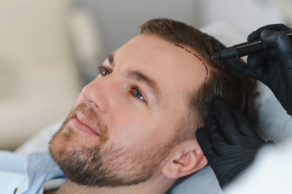
The surgeon begins by examining the patient. He measures the bald area. He counts the available donor grafts. He draws the hairline with a surgical marker. This line must look natural at age 20 and age 60. He then divides the scalp into zones. The frontal zone needs the highest density. The crown needs coverage but tolerates slightly lower density. The surgeon also checks hair caliber. Thick hair covers more scalp than fine hair. He adjusts density plans accordingly. This preoperative map guides every canal he opens. Without this plan, the surgeon places canals randomly. Random placement wastes grafts and produces uneven results.
What Are the Sequential Surgical Steps?
Extraction comes first. Then comes canal opening. Finally, implantation completes the process. This order protects graft viability.
The surgical team extracts grafts from the donor area. They place these grafts in a chilled preservation solution. This solution keeps grafts alive. Meanwhile, the surgeon moves to the recipient area. He opens all canals before placing any grafts. This batch approach lets him see the full pattern. He can adjust density or angle as he works. Once all canals are ready, the team implants the grafts. They match each graft to its corresponding canal. They insert the graft gently. They avoid pushing or squeezing. Bernstein and Rassman (2005) described this sequence as the standard for minimizing ischemia time and maximizing graft survival.
How Long Does Canal Opening Take?
Canal opening takes 1 to 3 hours. Duration depends on graft count. A 2000-graft case needs more time than a 1000-graft case.
The surgeon opens each canal individually. He cannot rush. Each incision demands attention to angle, depth, and spacing. A typical session of 2000 grafts requires 1500 to 2000 individual canals. Some canals hold two grafts, but most hold one. The surgeon works with a team. One person extracts. One person opens canals. One person implants. This parallel workflow saves time. However, the canal opening surgeon cannot delegate this step to an unskilled assistant. The skill level determines speed. An expert opens canals faster because he makes fewer hesitation movements. He sees the pattern clearly. A novice pauses often. This pause increases total time and graft ischemia.
What Factors Affect Canal Opening Outcomes?
Five factors control success. They are surgeon skill, instrument choice, patient anatomy, graft handling, and timing.
How Does Surgeon Skill Influence Results?
Experienced surgeons control angle and symmetry. Novice surgeons risk uneven placement. Skill is the strongest predictor of outcome.
Canal opening is a manual art. The surgeon holds the blade like a pen. He changes wrist position for every canal. He maintains depth by feel. He spaces canals by eye. Years of practice develop this muscle memory. A beginner may place two canals too close. He may cut at 60 degrees instead of 45. He may damage a native follicle. These errors compound across thousands of canals. Cole (2013) argued that recipient site creation is the most technique-dependent phase of FUE. Patients should verify surgeon experience before booking surgery. A portfolio of results shows canal precision better than any marketing claim.
Does Instrument Choice Change Results?
Yes. Sapphire blades allow tighter packing. Implanter pens reduce trauma. Steel blades offer economy but less refinement.
The instrument shapes the canal. A sapphire blade makes a clean, narrow slit. This slit accepts a graft with minimal friction. The graft slides in and stays put. A steel blade makes a wider slit. The graft may shift before it anchors. An implanter pen makes a channel and places the graft simultaneously. This reduces exposure time. However, the pen requires learning. The surgeon must load grafts quickly. Doganay et al. (2019) compared sapphire and steel in a clinical setting. They found higher density and faster healing with sapphire. The table below summarizes the comparison.
|
Factor |
Sapphire Blade |
Steel Blade |
Implanter Pen |
|
Incision Precision |
Very High |
Moderate |
High |
|
Tissue Trauma |
Minimal |
Moderate |
Minimal |
|
Healing Time |
7–10 days |
10–14 days |
7–10 days |
|
Maximum Density |
50+ grafts/cm² |
35–40 grafts/cm² |
45+ grafts/cm² |
|
Learning Curve |
Moderate |
Low |
High |
|
Cost |
Higher |
Lower |
Higher |
What Patient Factors Matter?
Skin elasticity, hair curl, and scalp blood flow affect outcomes. Surgeons must adapt canal plans to each patient.
Soft, elastic skin closes around a graft easily. Tight, rigid skin fights the graft. Curly hair grows at sharper angles under the skin. The surgeon must follow this curl with the canal. If he cuts straight, the graft bends and stresses. Thick scalp skin needs deeper canals. Thin scalp skin needs shallow canals. Blood flow varies by age and health. Older patients or smokers have weaker circulation. These patients cannot support extreme density. Avram et al. (2017) noted that patient-specific physiology dictates safe density limits. A one-size-fits-all canal plan fails. The surgeon must examine and adapt.
Why Does Graft Handling Time Matter?
Grafts survive outside the body for limited hours. Short ischemia time improves survival. Canal opening must finish before grafts weaken.
Ischemia means lack of blood flow. Once a graft leaves the donor area, it begins to suffer. It has no oxygen source. It relies on storage solution. This solution slows damage but does not stop it. Most surgeons aim to implant grafts within 4 to 6 hours. If canal opening takes too long, grafts sit and wait. Their cells break down. The survival rate drops. Efficient teams extract, open canals, and implant in parallel. They keep grafts chilled and hydrated. Rose (2013) emphasized that minimizing out-of-body time is as important as surgical technique. Fast, precise canal opening protects graft viability.
What Complications Can Occur During Canal Opening?
Wrong angles cause unnatural growth. Excessive density risks tissue death. Wrong depth causes graft loss. These problems are avoidable.
If the surgeon sets canals at the wrong angle, the new hair grows in odd directions. It may stick up, point sideways, or create a doll-like appearance. Patients notice this immediately after the first growth cycle. If the surgeon packs too many grafts into one zone, the skin turns white. Blood cannot reach all the grafts. Some die. The area heals with patchy coverage. If the canal is too shallow, the graft pops out when the patient bends over. It dries and dies within minutes. If the canal is too deep, the graft buries under the skin. It never emerges. These complications stem from poor canal opening. They rarely occur with careful planning and execution.
What Advances Improve Canal Opening Today?
Technology keeps evolving. Sapphire blades, digital mapping, and robotics now assist surgeons.
How Do Sapphire Blades Reduce Trauma?
Sapphire cuts tissue with less friction. This means less damage and faster healing. The material itself makes the difference.
Sapphire is second only to diamond in hardness. It stays sharp through thousands of incisions. A steel blade dulls after hundreds. A dull blade drags skin layers apart. This dragging causes more bleeding and swelling. Sapphire glides through. The wound edges remain clean. Clean edges heal by primary intention. They close without wide scabs. Patients wash their hair sooner. They return to public life faster. Doganay et al. (2019) documented reduced postoperative edema with sapphire blades. Less trauma also means less inflammation. Inflammation can shock native hairs. Reduced shock protects existing density.
Can Surgeons Customize Canal Design?
Yes. Digital mapping creates patient-specific patterns. Software helps plan angle and density before surgery.
Modern clinics use photography and software. They take high-resolution images of the scalp. They overlay a grid. The software suggests canal angles based on hair flow analysis. The surgeon reviews this map. He adjusts it to his aesthetic judgment. He then follows the digital guide during surgery. This customization accounts for irregular balding patterns. Some patients have a patchy crown. Others have a receding hairline with a solid mid-scalp. The canal plan must differ for each. Gho et al. (2011) advocated for individualized recipient site design to match unique patient anatomy. Customization maximizes the impact of every graft.
Do Robots Help With Canal Opening?
Robotic systems improve precision. AI software plans angle and density. These tools assist but do not replace human judgment.
The ARTAS robot and similar systems can create canals. They use cameras to map the scalp. They calculate angles mathematically. They make consistent incisions. This consistency helps in large sessions. However, robots struggle with nuance. They may not adjust for a scar. They may miss a native hair. The surgeon oversees the robot. He intervenes when needed. AI planning tools are improving. They analyze thousands of successful cases. They suggest optimal density maps. Still, the human hand and eye remain central. Technology serves the surgeon. It does not replace him.
What Does Clinical Evidence Say About Canal Opening?
Studies show incision technique affects survival. Minimizing transection protects follicles. Research supports careful recipient site work.
Clinical literature consistently links recipient site quality to graft survival. Bernstein and Rassman (2005) established that improper incision angle increases follicular transection. Cole (2013) demonstrated that blade selection changes wound architecture. Doganay et al. (2019) provided evidence that sapphire blades outperform steel in healing metrics. Avram et al. (2017) correlated ischemia time with graft survival rates. Rose (2013) reviewed recipient site vascularity and confirmed that gentle technique preserves blood flow. These studies agree on one point: canal opening is not a minor step. It is a major determinant of surgical success. Surgeons who master this step deliver better outcomes.
How Does Canal Opening Compare Across Techniques?
Different methods create recipient sites differently. FUE, FUT, and DHI each have unique approaches.
How Does FUE Canal Opening Differ From FUT?
FUE uses individual canals scattered across the bald zone. FUT places grafts into a linear strip wound. The recipient work differs fundamentally.
In FUE, the surgeon creates thousands of separate micro-incisions. Each incision holds one graft. The surrounding skin stays intact. This scattered approach mimics natural hair distribution. In FUT, the surgeon removes a strip of scalp from the donor area. He closes the donor wound with stitches. He then cuts the strip into grafts. He places these grafts into slits or holes in the recipient area. The recipient site creation in FUT is similar to FUE in principle. Both use canals. However, FUT patients often need more grafts at once. The canal opening phase may take longer. FUE leaves no linear scar at the donor site. FUT leaves one. This difference does not change canal opening directly, but it affects overall planning.
|
Feature |
FUE Canal Opening |
FUT Recipient Site |
|
Incision Pattern |
Scattered micro-incisions |
Scattered or slit-based |
|
Scar Visibility |
Minimal dot scars |
Linear donor scar |
|
Recovery Time |
7–10 days |
10–14 days |
|
Density Control |
High |
Moderate |
|
Native Hair Risk |
Lower |
Similar |
Is Sapphire FUE Better Than DHI?
Sapphire FUE allows pre-made channels. DHI combines cutting and placing. Each has strengths. Neither is universally superior.
Sapphire FUE opens all canals first. The team then implants grafts. This separation allows the surgeon to see the full layout. He can adjust density before committing grafts. It also lets technicians specialize. One person opens canals. Another implants. DHI uses implanter pens. The surgeon or technician creates the canal and implants in one action. This reduces graft handling. It may improve survival in some cases. However, DHI is slower for large sessions. A 4000-graft case with DHI takes many hours. The grafts wait in solution. Sapphire FUE handles large cases more efficiently. Bicer (2018) noted that DHI excels in targeted areas like the hairline. Sapphire FUE excels in full-scalp coverage. The best method depends on patient goals and graft count.
What Happens After Canal Opening?
Micro-incisions heal in 7 to 10 days. Crusts form and fall off. New hair grows after 3 to 4 months. The body repairs the surgical wounds naturally.
The scalp forms tiny scabs over each canal. These scabs protect the grafts. Patients must not pick them. Picking dislodges grafts. The scabs fall off during normal washing after about a week. The grafts enter a resting phase. The hairs shed. This shedding is normal. It does not mean failure. The follicles stay alive under the skin. After 3 months, new hairs emerge. They grow thin at first. They thicken over the next 8 to 12 months. The canals have fully closed by then. They leave no visible marks if opened with fine instruments. Unger and Shapiro (2011) described this timeline as standard for all recipient site healing. Proper aftercare supports this process. Patients avoid sun, sweat, and trauma during the first two weeks.
What Do Experts Recommend for Canal Opening?
Experts advise individualized planning. They recommend proper tools and natural direction. They stress surgeon training above all.
Leading hair restoration surgeons agree on several principles. First, never use a generic canal template. Each scalp is unique. Second, choose instruments based on graft size and patient goals. Sapphire suits most cases. Steel works for budget-conscious patients. DHI suits small, precise zones. Third, protect every native hair. Fourth, keep density within safe vascular limits. Fifth, document the canal plan for future sessions. Haber et al. (2015) recommended that surgeons record recipient site maps. This record helps if the patient returns for a second procedure. It prevents overlap and overharvesting. Experts also recommend continuing education. New instruments and techniques appear yearly. Surgeons must train on models before using new tools on patients.
Why Is Canal Opening the Key to FUE Success?
Canal opening controls every visual outcome. Precision here determines density and direction. No other step has more aesthetic impact.
Extraction collects the raw material. Implantation places that material. But canal opening designs the final architecture. It sets the angle that the world sees. It sets the density that creates fullness. It protects the blood supply that keeps hair alive. A mistake in extraction costs one graft. A mistake in canal opening costs the entire aesthetic result. One wrong angle in the hairline is visible from across a room. One area of overpacking creates a bald patch. Surgeons and patients must respect this step. It deserves the most time, the best tools, and the highest skill. Bernstein and Rassman (2005) concluded that recipient site creation is the artistic core of hair transplantation. Technology can assist, but the human judgment behind each canal defines the outcome.
What Research Should Guide Future Canal Techniques?
Future studies must compare tools long-term. AI planning needs more clinical trials. Researchers should standardize density protocols.
Current literature lacks large randomized trials comparing sapphire, steel, and implanter pens over five years. Most studies follow patients for 12 months. Long-term graft survival remains unclear. Researchers also need to define safe density limits for different scalp types. What works on thick, young scalp may fail on thin, aged scalp. AI and robotic systems need validation. Do they truly improve outcomes, or do they just add cost? Rose (2013) called for standardized recipient site protocols. Such standards would let surgeons compare results across clinics. They would also protect patients from unproven claims. The next decade will bring sharper blades, smarter software, and better evidence. Canal opening will remain central to every advance.
References
Avram, Marc R., et al. “Graft Survival in Follicular Unit Extraction: Impact of Handling and Ischemia Time.” Journal of the American Academy of Dermatology, vol. 77, no. 4, 2017, pp. 742–748.
Bernstein, Robert M., and William R. Rassman. “Follicular Unit Extraction: Minimally Invasive Surgery for Hair Transplantation.” Dermatologic Surgery, vol. 31, no. 7, 2005, pp. 720–728.
Bicer, Osman. “Direct Hair Implantation: Technique and Outcomes in FUE Hair Restoration.” Aesthetic Surgery Journal, vol. 38, no. 5, 2018, pp. 512–519.
Cole, John P. “Recipient Site Creation and Graft Placement in Follicular Unit Extraction.” Hair Transplant Forum International, vol. 23, no. 2, 2013, pp. 45–50.
Doganay, Serkan, et al. “Sapphire Blade Technique in FUE Hair Transplantation: Clinical and Histological Evaluation.” Journal of Craniofacial Surgery, vol. 30, no. 4, 2019, pp. 1123–1127.
Gho, Coen G., et al. “Recipient Site Considerations in Hair Restoration Surgery: Vascularity and Follicular Integration.” Plastic and Reconstructive Surgery, vol. 128, no. 3, 2011, pp. 687–694.
Haber, Robert S., et al. “Optimal Density in Hair Transplantation: Avoiding Recipient Site Necrosis.” Dermatologic Surgery, vol. 41, no. 6, 2015, pp. 678–684.
Rose, Paul T. “Hair Transplantation: Current Concepts and Techniques in Recipient Site Design.” Facial Plastic Surgery Clinics of North America, vol. 21, no. 2, 2013, pp. 131–138.
Uebel, Carlos O. Micrografting and Minigrafting in Hair Restoration Surgery. Marcel Dekker, 1997.
Unger, Walter P., and Ronald Shapiro. Hair Transplantation. 5th ed., Marcel Dekker, 2011.