Crown hair transplantationHair transplantation is a surgical procedure that involves the extraction of hair follicles from a designated donor site, followed by... restores hair to the vertex using advanced surgical techniques. Surgeons must reconstruct the natural whorl pattern while carefully managing limited donor grafts. Success depends on individualized planning, precise execution, and realistic patient expectations.
A crown hair transplant reverses baldness at the top-back portion of the head. This area displays a unique spiral pattern called the whorl. Crown restoration demands more precision than frontal hair transplant surgery because the whorl creates circular growth directions. Surgeons must rebuild this pattern to achieve natural results.
Androgenetic alopecia causes most crown hair loss. This condition shrinks hair follicles over time. The crown often thins faster than other areas. Patients notice a bald spot expanding from the vertex. Early intervention preserves donor hair for future sessions. Modern techniques like FUE, DHI, and Sapphire FUE allow surgeons to rebuild the crown with remarkable accuracy. Individualized planning remains essential because each patient presents a different whorl pattern, hair density, and balding progression.
What Is Crown Hair Loss and Why Does It Occur?
Crown hair loss is baldness at the vertex caused mainly by androgenetic alopecia. DHT shrinks follicles and creates a visible bald spot. The circular whorl pattern makes this area uniquely challenging to restore.
Crown hair loss affects the vertex region and creates a visible bald spot at the top-back of the head. This type of balding follows distinct patterns compared to frontal recession. The circular whorl complicates both diagnosis and treatment. Patients often notice crown thinning when hair parting reveals scalp visibility. Understanding the anatomy and causes helps patients make informed decisions about restoration.
What Is the Crown Area?
The crown area covers the vertex at the upper-back scalp. Hair here grows in a spiral whorl pattern that radiates outward from a central point. This radial growth differs sharply from the forward growth of frontal hair.
The crown area covers the vertex region at the upper-back scalp. This region forms a circular zone where hair radiates outward in a spiral formation. The anatomical term “vertex” describes this highest point at the back of the head.
The natural spiral and whorl pattern makes the crown unique. Most people display a single clockwise or counterclockwise whorl. Some individuals possess double whorls. Hair streams outward from a central point. Barbers and stylists must cut against this pattern carefully. The whorl creates natural volume and movement.
Crown hair differs significantly from frontal scalp hair. Frontal hair grows forward in a relatively straight direction. Crown hair radiates in multiple angles. This radial growth complicates transplantation. Surgeons must implant grafts at varying angles around a central point. Frontal restoration follows a linear framework. Crown restoration follows a circular framework. The frontal hairline frames the face. The crown frames the back profile. Both areas require distinct surgical approaches.
What Causes Crown Baldness?
Androgenetic alopecia causes the vast majority of crown baldness. Genetics, age, and hormones all contribute to progressive vertex thinning. Diffuse thinning can also spread evenly across the crown without forming a distinct bald patch.
Androgenetic alopecia causes the vast majority of crown baldness. This genetic condition affects both men and women. Dihydrotestosterone (DHT) attacks susceptible follicles. The hormone shrinks follicles progressively. Hair grows thinner and shorter. Eventually, follicles stop producing visible hair. The crown often shows the first noticeable thinning in male pattern baldness.
Hormonal and genetic factors drive this process. The AR gene regulates androgen receptors. Variations in this gene increase DHT sensitivity. Family history strongly predicts crown balding patterns. Fathers and grandfathers with vertex baldness indicate higher risk. Age-related thinning accelerates after forty. Hair density decreases naturally with time. The combination of genetics and aging creates visible crown loss.
Diffuse thinning represents another common pattern. Patients lose hair evenly across the crown. This pattern spreads progressively. The vertex loses coverage without a distinct bald patch. Progressive vertex loss continues without treatment. Medical therapy can slow this expansion. Transplantation becomes necessary when medication fails.
Why Is the Crown Difficult to Restore?
Circular growth directions, light reflection on the curved scalp, and the large surface area make crown restoration complex. Surgeons must place every graft at a precise radial angle. Ongoing balding progression also complicates long-term planning.
Circular hair growth directions create the primary challenge. Surgeons must place each graft at a precise radial angle. A single incorrect angle disrupts the entire whorl. The circular pattern requires 360-degree planning. Frontal restoration uses primarily forward angles. Crown restoration uses angles that shift every few millimeters.
Light reflection creates an illusion of reduced density at the crown. Overhead lighting exposes the scalp more easily at the vertex. The curved surface reflects light directly. Even healthy crowns appear thinner under bright light. Transplanted hair must achieve higher density to overcome this optical effect.
The crown covers a larger surface area than patients expect. The vertex region extends several centimeters in all directions. Complete restoration often requires two thousand to three thousand grafts. Larger bald areas demand even more. The ongoing progression of crown balding complicates planning. Surgeons must reserve donor grafts for future sessions. Young patients may lose more native hair around the transplant. Strategic distribution prevents overuse of limited donor supply.
Who Qualifies for Crown Hair Transplant and How Do Surgeons Evaluate Candidates?
Ideal candidates have stable donor density, realistic expectations, and isolated crown thinning. Surgeons assess age, Norwood stage, hair caliber, and future loss risk. Donor areaThe Source of Restoration The donor area plays a critical role in hair transplantation, as it serves as the source... management protects long-term results.
Surgeons evaluate candidates through physical examination, medical history, and long-term planning. The crown requires careful candidate selection because graft usage is high and future balding may progress. Not every patient with crown thinning qualifies immediately. Surgeons assess donor reserves, hair characteristics, and realistic goals before approving surgery.
What Makes an Ideal Candidate for Crown Hair Transplant?
An ideal candidate possesses a stable donor area with sufficient density and maintains realistic expectations about coverage limits. Patients with isolated crown thinning and strong surrounding hair make the best candidates. Thick, curly hair provides better visual coverage per graft.
Stable donor area represents the first requirement. The back and sides of the scalp must maintain dense, healthy hair. Surgeons examine donor density under magnification. Patients with thick donor regions achieve better crown coverage. Thin or unstable donor areas limit graft availability.
Sufficient donor density determines coverage potential. Patients need enough grafts for the crown and future needs. Young patients with early crown loss require conservative planning. Surgeons must preserve grafts for progressive balding.
Realistic expectations separate good candidates from poor ones. Crown transplants rarely match native density. Patients must understand the “illusion of fullness” principle. Complete baldness cannot transform into teenage density. Improvement matters more than perfection.
Patients with isolated crown thinning make excellent candidates. These individuals maintain strong frontal hairlines. The crown represents their only balding zone. Targeted restoration solves their primary concern. Surgeons can focus all grafts on one region.
What Factors Affect Suitability for Crown Restoration?
Age, Norwood classification, hair caliber, scalp contrast, and existing miniaturized hairs all affect candidacy. Young patients need conservative planning to preserve grafts for future loss. High color contrast demands higher density for acceptable visual results.
Age and future hair loss progression dominate the evaluation. Surgeons hesitate to transplant crowns in patients under twenty-five. Early intervention risks misallocation of grafts. Young patients may develop extensive baldness later. Surgeons prefer patients with stabilized loss patterns.
Norwood scale classification guides surgical decisions. The Norwood system measures male pattern baldness stages. Patients at stage three vertex or higher qualify for crown work. Advanced stages require comprehensive planning. Surgeons must address multiple zones across multiple sessions.
Hair caliber and scalp contrast influence visual results. Thick hair shafts create better coverage. Each thick graft blocks more scalp visibility. Dark hair on light skin shows more contrast. This contrast demands higher density for acceptable appearance. Curly hair provides excellent coverage. Each curl occupies more space visually.
Existing miniaturized hairs complicate crown restoration. These thin, weak hairs surround the bald area. Surgeons must work around them carefully. Implantation can damage miniaturized neighbors. Strategic placement preserves these native hairs.
Why Does Donor Area Management Matter for Crown Transplants?
The safe donor zone provides DHT-resistant grafts, but overharvesting destroys this resource permanently. Surgeons must calculate lifetime graft supply and reserve follicles for future sessions. Conservative extraction preserves cosmetic balance at the back and sides.
The safe donor zone concept protects long-term results. This zone sits at the back and sides of the scalp. Hair in this region resists DHT naturally. Surgeons extract grafts exclusively from this zone. Overharvesting damages the donor area permanently. Visible thinning at the back destroys cosmetic balance.
Long-term graft conservation requires discipline. Surgeons calculate lifetime graft supply. A typical donor area contains six thousand to eight thousand viable grafts. Crown restoration may consume one-third of this supply. Patients with progressive balding need grafts for future hairline work. Conservative crown planning preserves options.
Planning for future sessions defines responsible surgery. Young patients often need multiple procedures over decades. Surgeons must anticipate this need. They distribute grafts strategically across time. The crown receives enough grafts for current improvement. Surgeons reserve remaining grafts for later expansion.
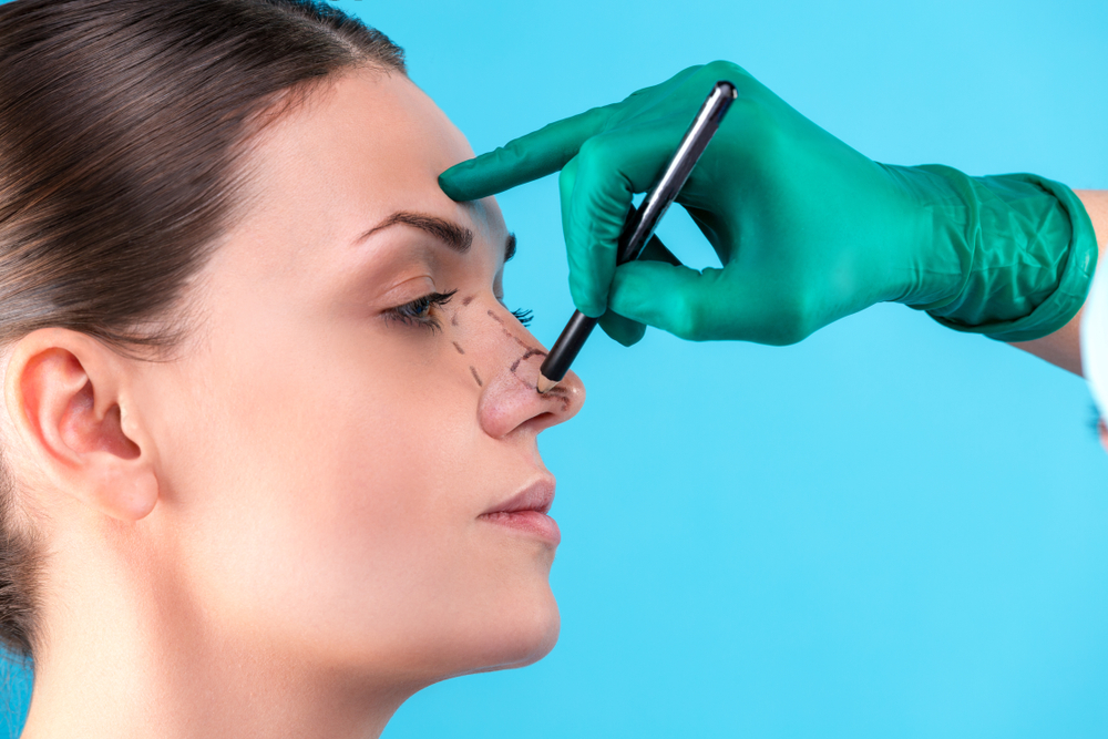
How Do Surgeons Assess and Design the Crown Before Surgery?
Preoperative assessment includes density measurement, hair shaft analysis, whorl mapping, and mathematical graft planning. Surgeons photograph and mark the scalp to guide incision angles. Each patient receives a unique surgical map based on individual anatomy.
Preoperative assessment combines physical measurement, pattern mapping, and mathematical planning. Surgeons must understand the exact whorl structure before making any incisions. They analyze hair density, thickness, and curl. They photograph and mark the scalp. This preparation ensures the transplant follows the patient’s unique anatomy.
How Do Surgeons Analyze Scalp and Hair Before Crown Surgery?
Surgeons measure follicular unit density, hair shaft thickness, scalp elasticity, and curl characteristics. Thick shafts above eighty micrometers block more scalp visibility. These measurements determine graft selection and placement strategy.
Density measurements establish baseline data. Surgeons count follicular units per square centimeter. Normal density ranges from seventy to one hundred units. Crown restoration targets lower densities than native scalp. Patients with higher donor density achieve better results.
Hair shaft thickness determines coverage efficiency. Thick shafts block more light and scalp visibility. Surgeons measure diameter in micrometers. Coarse hair above eighty micrometers provides excellent coverage. Fine hair below fifty micrometers requires more grafts for similar visual density.
Scalp elasticity affects surgical approach. Loose scalps allow easier extraction and implantation. Tight scalps limit surgical flexibility. Surgeons assess elasticity during consultation.
Hair curl characteristics influence implantation strategy. Curly hair grows in unpredictable directions. Surgeons must account for curl pattern when placing grafts. Straight hair follows more predictable paths. Wavy hair falls between these extremes.
How Do Surgeons Map the Crown Whorl?
Surgeons identify whether the whorl runs clockwise or counterclockwise and note single or double patterns. They mark the central point and radiating lines on the scalp before surgery. Accurate mapping prevents unnatural growth angles.
Clockwise patterns appear in approximately eighty percent of patients. Counterclockwise whorls appear in the remaining twenty percent. Surgeons identify the pattern before making incisions. Incorrect identification leads to unnatural growth.
Single whorls dominate most scalps. Double whorls create complex patterns. Some patients display irregular or disrupted whorls. Surgeons must map these variations precisely. They mark the central point and radiating lines.
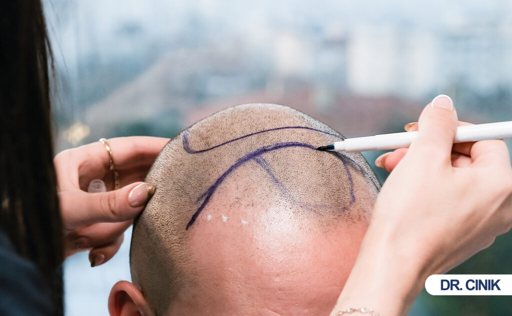
Personalized scalp mapping ensures accurate reconstruction. Surgeons draw the whorl pattern on the scalp before surgery. They photograph the area from multiple angles. These markings guide incision angles during the procedure. Each patient receives a unique map. No two crown patterns match exactly.
What Principles Guide Surgical Planning for the Crown?
Surgeons calculate surface area and multiply by target density of thirty to forty grafts per square centimeter. They preserve natural radial direction and plan for future balding progression. Strategic spacing prevents the “donut” effect as native hair continues to thin.
Determining graft distribution requires mathematical precision. Surgeons calculate the crown surface area. They multiply this area by target density. Typical crown targets range from thirty to forty grafts per square centimeter. Frontal areas often receive higher densities. The crown accepts slightly lower numbers due to whorl complexity.
Preserving natural hair direction remains critical. Surgeons must match existing growth patterns. They follow the radial lines of the whorl. Each incision points toward the central swirl. Deviations create messy, unnatural appearance.
Planning for future balding progression protects long-term aesthetics. Surgeons leave space for future loss. They avoid placing grafts too densely at the edges. Progressive baldness may create a “donut” effect around the transplant. Strategic spacing prevents this problem.
What Techniques Do Surgeons Use for Crown Hair Transplant?
Surgeons primarily use FUE, DHI, and Sapphire FUE for crown restoration. Each technique offers specific advantages for whorl reconstruction and dense packing. Technique selection depends on patient goals, donor characteristics, and surgeon expertise.
Surgeons employ multiple techniques for crown restoration. Each method offers specific advantages for whorl reconstruction. Technique selection depends on patient goals, donor characteristics, and surgeon expertise. FUE, DHI, and Sapphire FUE dominate modern crown work. Each approach offers distinct benefits for whorl precision and density.
How Does FUE Crown Hair Transplant Work?
FUE removes individual grafts with micro-punches and allows selective harvesting for crown density. Surgeons create spiral recipient sites that match the natural whorl. This technique leaves tiny dot scars and provides excellent angle control.
Follicular unit extraction methodology removes individual grafts. Surgeons use micro-punches to extract follicles. Each punch measures between 0.7 and 1.0 millimeters. The procedure leaves tiny dot scars. These scars hide within surrounding hair.
FUE offers distinct advantages in crown restoration. Surgeons select grafts with specific hair counts. They choose multi-hair grafts for central density. They place single-hair grafts at the edges. This selection creates natural density variation.
Precision implantation follows spiral patterns. Surgeons create recipient sites with custom angles. They work outward from the whorl center. Each site matches the natural radial direction. FUE allows flexible graft placement across the entire crown.
How Does DHI Crown Hair Transplant Differ?
DHI uses Choi implanter pens to insert grafts directly without pre-making recipient sites. This method enables denser packing and precise angle control. It reduces graft handling time and minimizes trauma to surrounding tissue.
Direct implantation approach eliminates recipient site creation first. Surgeons load grafts into Choi implanter pens. These pens insert grafts directly into the scalp. The technique combines incision and implantation into one step.
Choi implanter pen usage requires specialized training. Surgeons hold the pen like a writing instrument. They press the graft into the scalp at controlled depth. The pen releases the follicle smoothly. This method reduces handling time.
DHI benefits crown work through dense packing capabilities. Surgeons place grafts closer together. The technique minimizes trauma to surrounding tissue. Angle control improves because the pen guides direction precisely. Crown restoration benefits from these dense, accurate placements.
What Role Does Sapphire FUE Play in Crown Restoration?
Sapphire blades create finer, cleaner incisions than steel blades. Smaller cuts heal faster and reduce postoperative crusting. Improved accuracy helps surgeons reconstruct the whorl with exceptional precision.
Sapphire bladeA Modern Innovation in Hair Transplants The sapphire blade is an advanced tool used to create precise incisions during hair... technology replaces steel blades. Surgeons use blades made from synthetic sapphire. These blades stay sharper longer. They create cleaner, more precise incisions.
Smaller incisions improve healing significantly. Sapphire blades make finer cuts than steel. These micro-incisions close faster. Patients experience less postoperative crusting. The crown area heals more uniformly.
Improved graft placement accuracy enhances whorl reconstruction. Surgeons control depth and angle more precisely. The sharp blade enters the scalp smoothly. Grafts fit tightly into these refined sites. Crown patterns require this level of precision.
How Do DHI and Sapphire FUE Compare for Crown Restoration?
DHI and Sapphire FUE represent advanced implantation techniques that improve upon standard FUE. Both methods use the same follicular unit extraction from the donor area. The difference lies in recipient site creation and graft placement.
DHI excels at dense packing and angle precision. The Choi implanter pen inserts grafts directly without pre-making sites. This reduces graft handling time and allows tighter placement. DHI suits patients needing maximum crown density. The technique demands specialized training and a larger team.
Sapphire FUE focuses on incision quality and healing speed. The synthetic sapphire blade creates finer, sharper cuts than steel. These micro-incisions close faster and produce less crusting. Sapphire FUE benefits patients with sensitive scalps or those wanting quicker recovery. The blade allows precise depth and angle control for whorl reconstruction.
Both techniques avoid linear scars. Both achieve natural results when executed properly. Surgeons may combine elements of both approaches. Some use sapphire blades to create sites and standard forceps for placement. Others use DHI pens throughout. Technique choice depends on patient goals, scalp characteristics, and surgeon expertise.
|
Feature |
DHI |
Sapphire FUE |
|
Site creation |
Simultaneous with implantation |
Pre-made with sapphire blade |
|
Incision size |
Pen tip |
Ultra-fine sapphire blade |
|
Dense packing |
Excellent |
Very good |
|
Healing time |
Standard |
Faster due to finer cuts |
|
Crusting |
Moderate |
Minimal |
|
Training requirement |
Specialized |
Advanced |
|
Crown whorl precision |
High |
Very high |
How Do Surgeons Calculate Grafts for Crown Hair Transplant?
Graft numbers depend on crown size, hair characteristics, and target density. Small thinning needs one thousand to one thousand five hundred grafts. Extensive baldness may require two thousand five hundred to four thousand grafts across multiple sessions.
Graft calculation determines the success and naturalness of crown restoration. Surgeons measure the bald area and set density targets. They balance patient desires against donor limits. The crown often requires more grafts than patients expect.
How Many Grafts Does a Crown Hair Transplant Require?
Small cases need one thousand to one thousand five hundred grafts. Moderate vertex baldness requires one thousand five hundred to two thousand five hundred grafts. Extensive baldness demands two thousand five hundred to four thousand grafts.
Small crown thinning cases need one thousand to one thousand five hundred grafts. These patients display early vertex loss. The bald area measures less than five centimeters across. Strategic placement creates significant visual improvement.
Moderate vertex baldness requires one thousand five hundred to two thousand five hundred grafts. The bald patch expands visibly. Patients notice the crown under normal lighting. This stage represents the most common presentation.
Extensive crown baldness demands two thousand five hundred to four thousand grafts. The vertex shows complete or near-complete baldness. Surrounding areas also thin significantly. These cases may need multiple sessions. Surgeons must balance crown coverage against donor preservation.
|
Severity |
Graft Range |
Expected Coverage |
|
Small thinning |
1,000–1,500 |
Early vertex fill |
|
Moderate baldness |
1,500–2,500 |
Full vertex coverage |
|
Extensive baldness |
2,500–4,000 |
Maximum coverage possible |
What Density Does the Crown Require?
Crown targets typically reach thirty to forty grafts per square centimeter. Native density ranges from seventy to one hundred, so transplants rely on the illusion of fullness. Strategic placement and multi-hair grafts create visual coverage without matching native numbers.
Typical FU/cm² targets range from thirty to forty grafts. Native scalp density reaches seventy to one hundred. Crown transplants intentionally use lower numbers. Surgeons distribute grafts strategically to create fullness illusion.
The crown appears thinner due to light reflection. The curved vertex surface catches overhead light. Transplanted hair must overcome this optical challenge. Higher density at the center helps. Lower density at the edges blends with surrounding hair.
The “illusion of fullness” principle guides crown planning. Surgeons do not replicate native density. They create visual coverage through strategic placement. Multi-hair grafts at the center block more light. Proper angulation creates overlapping shadows. The eye perceives density where actual numbers remain modest.
What Factors Influence Graft Numbers for Crown Restoration?
Crown size, hair thickness, color contrast, curl pattern, and donor availability all influence graft requirements. Thick, straight hair on light skin needs more grafts than curly, dark hair. Surgeons must balance patient desires against long-term donor preservation.
Crown size directly determines graft requirements. Larger bald areas need more coverage. Surgeons measure the bald zone in square centimeters. They multiply area by target density.
Hair thickness and color contrast affect visual density. Thick, dark hair on light skin needs more grafts. Thin, light hair on dark skin needs fewer grafts. Curly hair provides better coverage per graft. Straight hair requires higher graft counts.
Desired density sets patient expectations. Some patients want maximum possible coverage. Others accept moderate improvement. Surgeons align graft numbers with patient goals.
Donor availability limits all plans. Patients with low donor density cannot receive unlimited grafts. Surgeons must balance crown work against future needs. Conservative planning preserves donor resources.
What Happens During Crown Hair Transplant Surgery Step by Step?
Surgery involves anesthesiaEnsuring Comfort During Hair Transplants Sedation is used in hair transplantation to help patients remain calm and comfortable throughout the..., shaving, graft extraction, recipient site creation, and implantation. Surgeons work outward from the whorl center using radial incision patterns. The procedure typically lasts six to eight hours.
Crown transplantation follows a strict sequence. Surgeons prepare the scalp, extract grafts, create sites, and implant follicles. Each phase requires specific skills for crown work.
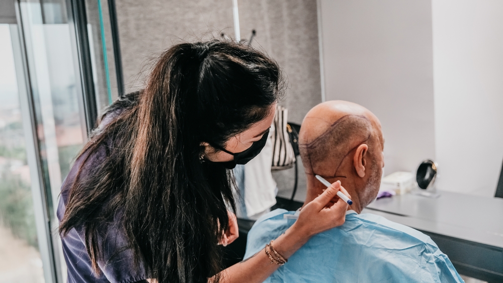
How Do Surgeons Prepare the Crown for Transplantation?
Surgeons shave the crown, administer local anesthesia, and mark the whorl pattern with surgical pens. Nerve blocks numb the entire vertex region. Clear markings guide precise angle and depth throughout the procedure.
Preoperative shaving allows clear access. Most patients shave the entire crown area. Some clinics offer unshaven FUE options. Shaved crowns provide better visibility for whorl mapping.
Anesthesia administration ensures comfort. Surgeons inject local anesthetic around the crown. Nerve blocks numb the entire vertex region. Patients remain awake throughout. They feel no pain during extraction or implantation.
Surgical markings guide the procedure. Surgeons draw the whorl pattern with surgical pens. They mark the central point clearly. They outline the bald zone boundaries. These markings remain visible throughout surgery.
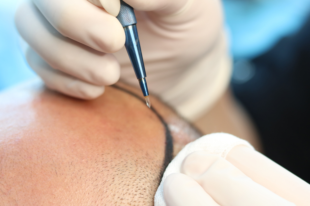
How Does Graft Extraction Proceed During Crown Surgery?
Surgeons extract grafts individually from the safe donor zone using standard FUE methodology. Technicians sort grafts by hair count immediately. Chilled holding solution preserves follicle viability regardless of whether implantation will use standard FUE, DHI, or Sapphire FUE channels.
Donor harvesting follows standard FUE protocols for all crown techniques. Surgeons extract grafts individually across the safe donor zone. They select grafts with appropriate hair counts. Technicians sort grafts by size immediately.
Graft preservation techniques maintain viability. Technicians place extracted follicles in chilled holding solution. This solution mimics physiological conditions. Temperature control slows metabolic activity. Grafts remain viable for several hours.
Minimizing follicular trauma protects graft quality. Surgeons align punches with hair angle. Incorrect alignment cuts follicles. Technicians handle grafts with fine forceps. They avoid crushing or drying the tissue. Each graft represents a precious, non-renewable resource.
How Do Surgeons Create Recipient Sites at the Crown?
Surgeons make incisions at thirty to forty-five degree angles radiating from the whorl center. Depth reaches four to six millimeters to protect blood supply. Each site follows the personalized scalp map to recreate natural orientation.
Angle and depth control demands constant attention. Crown incisions range from thirty to forty-five degrees. Depth reaches approximately four to six millimeters. Too shallow risks graft popping. Too deep damages blood supply.
Radial incision patterns recreate the whorl. Surgeons start at the central point. They create sites radiating outward. Each ring of sites follows the circular pattern. They adjust angles as they move around the swirl.
Recreating natural whorl orientation requires artistic skill. Surgeons must visualize the final growth pattern. They follow the patient’s original whorl direction. Double whorls need complex interweaving. Single whorls follow simpler radial lines.
How Does Graft Implantation Work at the Crown?
Surgeons place single-hair grafts at the edges and multi-hair grafts centrally for natural density variation. They build coverage through density layering in multiple passes. Strategic clustering mimics natural growth irregularity.
Single vs multi-hair graft placement follows strategic rules. Surgeons place single-hair grafts at the edges. These create soft, natural borders. They place two and three-hair grafts centrally. These provide density and coverage.
Density layering techniques build visual fullness. Surgeons implant grafts in multiple passes. They create a foundation layer first. They add density layers above. This stacking creates depth perception.
Strategic placement optimizes natural appearance. Surgeons avoid uniform distribution. They cluster grafts where needed. They leave slight gaps where native hair remains. This irregularity mimics natural growth patterns.
What Does Recovery Involve After Crown Hair Transplant?
Recovery includes immediate swelling, a temporary shedding phase, and gradual regrowth over twelve to eighteen months. Patients follow specific washing and activity protocols. PRP and medication support enhance healing and growth.
Recovery after crown transplant follows a predictable pattern. Patients experience immediate postoperative changes, a shedding phase, and gradual regrowth. Proper care protects graft survival and speeds healing.
What Happens Immediately After Crown Hair Transplant?
Swelling and redness peak within twenty-four hours and resolve in three to five days. Crusts form over graft sites and must remain undisturbed for seven to ten days. Patients sleep with their head elevated to protect new follicles.
Swelling and redness appear within twenty-four hours. The crown area becomes puffy. Fluid shifts downward due to gravity. Some patients notice forehead swelling. This resolves within three to five days.
Crusting and scab formation protect graft sites. Small blood clots seal each incision. These crusts remain for seven to ten days. Patients must not pick or scratch them. Premature removal dislodges grafts.
Sleeping position recommendations protect new grafts. Patients should sleep with their head elevated. They avoid touching the crown against pillows. Some clinics provide neck pillows. Back sleeping prevents pressure on the vertex.
What Is the Shedding Phase After Crown Transplant?
Temporary graft shedding begins around week two as hairs fall out while follicles remain anchored. Native shock loss may affect surrounding miniaturized hairs. New growth typically starts by month three.
Temporary graft shedding begins around week two. Newly implanted hairs fall out. The follicles remain anchored beneath the skin. This shock shedding alarms patients. It represents a normal, expected process.
Native hair shock loss affects surrounding hairs. Surgical trauma stresses nearby follicles. Some miniaturized hairs fall out. These may or may not return. PRP therapy reduces this risk.
Regrowth begins around month three. Tiny new hairs emerge from transplanted follicles. These hairs start thin and colorless. They gradually thicken and pigment. The crown shows early stubble by month four.
What Timeline Should Crown Transplant Patients Expect?
Early growth appears at three months, visible improvement shows by six months, and final maturation completes at twelve to eighteen months. Transplanted hair reaches full thickness and texture by the end of the first year.
Three-month early growth brings first visible changes. Patients notice fine hairs sprouting. These hairs lack full thickness. They appear soft and light-colored.
Six-month visible improvement shows meaningful coverage. Hairs gain length and diameter. The crown looks less bald under normal light. Patients can style hair with more confidence.
Twelve to eighteen month final maturation completes the process. Transplanted hair reaches full thickness. The texture matches native hair. The crown achieves maximum visual density. Patients evaluate final results at this stage.
What Postoperative Care Instructions Apply to Crown Transplants?
Patients begin gentle washing on day three and avoid heavy lifting for two weeks. Surgeons prescribe antibiotics and anti-inflammatory medication. PRP injections at one, three, and six months accelerate growth.
Washing protocol starts around day three. Patients gently rinse the crown with prescribed shampoo. They avoid direct water pressure. They pat dry without rubbing. Regular washing prevents infection and crust buildup.
Physical activity restrictions last two weeks. Patients avoid heavy lifting and straining. These activities increase blood pressure. Elevated pressure risks graft dislodgement. Light walking remains acceptable.
Medication and PRP support enhance results. Surgeons prescribe antibiotics for five days. Anti-inflammatory medication reduces swelling. PRP injections at one, three, and six months accelerate growth. Some patients continue finasteride or minoxidil.
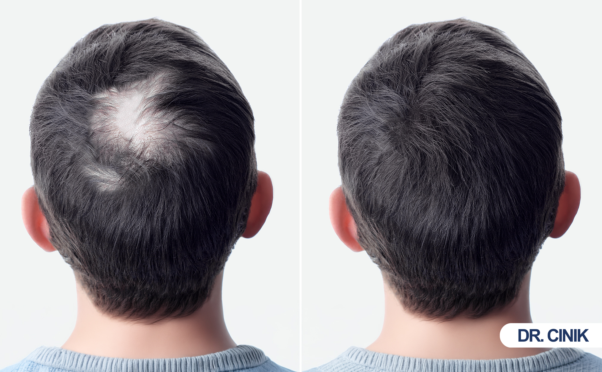
What Results and Long-Term Outcomes Can Patients Expect?
Crown transplants achieve sixty to seventy percent of native density with high naturalness when executed properly. Transplanted follicles last permanently due to donor dominance. Progressive surrounding loss may require future touch-ups.
Crown transplant results develop gradually. Patients must judge success by coverage improvement rather than perfect density. Long-term outcomes depend on surgical skill and ongoing hair preservation.
What Density and Coverage Can Crown Transplant Patients Expect?
Patients achieve sixty to seventy percent of native density, which creates acceptable visual coverage. Strategic placement matters more than maximum graft count. The illusion of fullness principle guides realistic outcomes.
Realistic crown density expectations remain modest. Crown transplants achieve sixty to seventy percent of native density. This percentage creates acceptable visual coverage. Patients must not expect complete restoration.
Crowns rarely achieve native density for several reasons. The large surface area dilutes graft impact. Light reflection exaggerates thinness. Limited donor supply prevents dense packing. Surgeons prioritize natural appearance over maximum numbers.
Visual fullness differs from actual density. Strategic placement creates perception of density. Proper angulation and layering matter more than graft count. A well-executed moderate-density transplant outperforms a poorly executed high-density one.
How Natural Do Crown Transplants Appear?
Naturalness depends entirely on directional accuracy and whorl recreation. Experienced surgeons replicate the central swirl and blend edges seamlessly. Poor angle placement creates chaotic, obviously artificial growth.
Directional accuracy determines naturalness. Each hair must follow the whorl pattern. Incorrect angles create messy, chaotic growth. Proper angles create seamless integration.
Whorl recreation success depends on surgical artistry. Experienced surgeons study the original pattern. They replicate the central swirl precisely. They blend edges with surrounding hair. The best crown transplants look indistinguishable from natural growth.
How Long Do Crown Hair Transplants Last?
Transplanted follicles maintain DHT resistance and grow permanently. Ninety percent graft survival represents standard outcomes. However, surrounding native hair may continue thinning and require medical management.
Donor dominance theory explains longevity. Hair from the safe donor zone resists DHT. These follicles maintain this resistance after transplantation. They continue growing for decades.
Stability of transplanted follicles remains high. Most grafts survive permanently. Ninety percent survival rates represent standard outcomes. Transplanted hair behaves like donor area hair.
Progressive surrounding hair loss creates future challenges. Native hair around the transplant may continue thinning. This process leaves the transplant isolated. Patients may need medical therapy to preserve surrounding hair. Future touch-up sessions address continued loss.
How Do Surgeons Assess Before and After Results?
Surgeons use standardized photography, trichoscopy, and patient satisfaction surveys. Consistent lighting and angles allow objective comparison. Density evaluation occurs microscopically and visually.
Patient satisfaction surveys measure subjective success. Patients rate density, naturalness, and overall improvement. High satisfaction correlates with realistic expectations.
Density evaluation uses standardized photography. Surgeons photograph the crown under consistent lighting. They compare preoperative and postoperative images. Trichoscopy provides microscopic density counts.
Photographic comparison standards ensure objective assessment. Surgeons use the same angles, lighting, and zoom. They capture vertex views consistently. These images document improvement for medical records.
What Risks and Complications Can Crown Transplants Cause?
Common side effects include temporary edema, folliculitis, numbness, and shock loss. Serious complications such as poor graft survival, overharvesting, and cobblestoning remain rare with experienced surgeons. Incorrect planning causes most failures.
All surgical procedures carry risks. Crown transplantation presents specific complications related to the unique anatomy. Most side effects resolve quickly. Serious complications remain rare with experienced surgeons.
What Are the Common Side Effects of Crown Transplant?
Edema peaks at day three and resolves within a week. Folliculitis responds to topical antibiotics. Temporary numbness fades in three to six months. Shock loss affects native hairs but most return.
Edema affects most patients temporarily. Fluid accumulates in the forehead and around the eyes. This swelling peaks at day three. It resolves without intervention within a week.
Folliculitis develops in some patients. Inflamed hair follicles create small pimples. This condition responds to topical antibiotics. Proper washing prevents most cases.
Temporary numbness occurs around the crown. Nerve endings require time to regenerate. Sensation returns within three to six months. Permanent numbness remains rare.
Shock loss affects native hairs near transplant sites. These hairs may shed temporarily. Most return within three months. Miniaturized hairs may not recover.
What Surgical Complications Can Occur?
Poor graft survival, unnatural growth direction, overharvesting, and cobblestoning represent potential complications. Survival rates below eighty percent indicate technical problems. Correct depth and angle control eliminate most risks.
Poor graft survival wastes donor resources. Dehydration, trauma, or improper handling kills follicles. Survival rates below eighty percent indicate problems. Experienced surgeons maintain ninety percent or higher.
Unnatural growth direction creates visual problems. Incorrect angles make hair stick up or lie flat. Whorl errors produce swirling chaos. Revision surgery becomes difficult.
Overharvesting damages the donor area permanently. Excessive extraction creates visible thinning. Patients cannot reverse this damage. Conservative harvesting prevents this tragedy.
Cobblestoning creates bumpy scalp texture. This complication occurs with improper depth control. Grafts sit too high or too deep. Correct technique eliminates this risk.
Why Do Some Crown Hair Transplants Fail?
Incorrect angle placement, insufficient graft numbers, poor donor planning, and unrealistic expectations cause most failures. Each wrong angle compounds whorl distortion. Underpowered transplants often look worse than untreated baldness.
Incorrect angle placement represents the leading cause. Surgeons unfamiliar with crown patterns make systematic errors. Each wrong angle compounds the problem. The entire whorl appears distorted.
Insufficient graft numbers create sparse coverage. Patients demand coverage beyond donor supply. Surgeons must refuse unrealistic requests. Underpowered transplants look worse than baldness.
Poor donor planning exhausts graft reserves. Aggressive crown work leaves nothing for future needs. Patients regret this allocation later. Conservative strategy serves better long-term.
Unrealistic expectations doom patient satisfaction. Some patients expect miracles. They ignore surgical limitations. Proper consultation prevents disappointment.
How Does Crown Transplant Differ from Hairline Transplant?
Crown restoration follows circular spiral patterns while hairline work follows linear forward growth. The crown requires more grafts for equivalent visual impact. Surgeons usually prioritize the hairline for younger patients.
Crown and hairline restoration serve different cosmetic purposes. They require distinct surgical strategies. Understanding these differences helps patients prioritize their goals.
What Are the Key Anatomical Differences?
Crown hair radiates in a spiral whorl while frontal hair grows forward in straight lines. The hairline frames the face directly and draws immediate attention. The crown remains less visible in frontal conversation but shows clearly from above.
Linear vs spiral growth defines the difference. Hairlines grow forward in relatively straight lines. Crowns grow outward in circular patterns. Surgeons must think linearly at the front. They must think radially at the back.
Visibility and cosmetic framing differ significantly. The hairline frames the face directly. People notice hairline problems immediately. The crown remains less visible in daily interaction. However, crown baldness shows clearly from above and behind.
How Do Density and Graft Distribution Differ Between Crown and Hairline?
The crown’s circular surface area demands more grafts than the linear hairline. Frontal grafts create dramatic framing effects with fewer follicles. Crown grafts spread across a wider zone with less impact per graft.
Crowns require more grafts for equivalent coverage. The circular surface area exceeds linear hairline length. Each square centimeter of crown needs careful attention. Hairline work concentrates grafts along a narrow band.
Coverage efficiency comparison favors the hairline. Frontal grafts create dramatic framing effects. Crown grafts spread across a wider zone. The impact per graft feels less dramatic at the vertex.
Which Area Should Surgeons Prioritize?
Surgeons typically prioritize the hairline because it frames the face and restores youthfulness. Young patients with progressive loss should delay crown work to preserve grafts. Crown restoration suits patients with stable hairlines.
Strategic restoration planning addresses this question. Surgeons typically prioritize the hairline first. The hairline frames the face and creates youthfulness. Crown work follows as a secondary procedure.
Younger patients with future balding progression need special consideration. Surgeons may delay crown work entirely. They preserve grafts for hairline maintenance. Crown restoration becomes appropriate when hairline stabilizes. Some patients choose crown work first if the hairline remains strong.
How Can Patients Enhance Crown Hair Transplant Results?
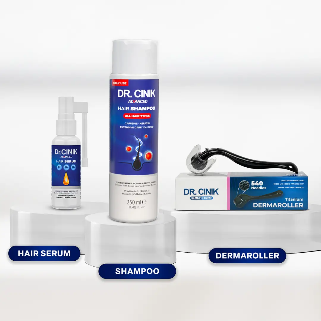
PRP therapy, medical treatments, and lifestyle changes all improve outcomes. Minoxidil, finasteride, and dutasteride protect native hair. Smoking cessation and proper nutrition support graft survival.
Medical and lifestyle support maximizes crown transplant outcomes. Patients should combine surgery with ongoing therapy. These adjuncts protect native hair and accelerate transplanted growth.
How Can PRP Therapy Enhance Crown Results?
PRP injections deliver concentrated growth factors that accelerate healing and reduce shedding. Khatu et al. (2014) showed significant density improvement with PRP. Regular sessions maintain long-term follicle health.
Platelet-rich plasma concentrates growth factors. Surgeons draw patient blood and spin it in centrifuges. The plasma layer contains platelets and cytokines. They inject this concentrate into the crown.
Healing acceleration benefits crown patients. PRP reduces inflammation faster. It speeds graft anchoring. Patients experience less shedding. Growth begins earlier and stronger.
Studies support PRP efficacy. Khatu et al. (2014) demonstrated significant improvement in hair density with PRP treatment. The growth factors stimulate follicle stem cells. Regular PRP sessions maintain results long-term.
What Medical Treatments Support Crown Transplants?
Minoxidil extends the growth phase, finasteride blocks DHT conversion, and dutasteride provides stronger suppression. These medications protect surrounding native hair. Patients often continue them indefinitely after surgery.
Minoxidil stimulates blood flow to follicles. This topical medication extends the growth phase. Patients apply it daily to the crown. It supports both native and transplanted hair.
Finasteride blocks DHT conversion. This oral medication slows progressive balding. It protects surrounding native hair. Patients often continue finasteride after transplantation.
Dutasteride provides stronger DHT suppression. This medication blocks both type 1 and type 2 enzymes. It suits patients with aggressive hair loss. Physicians monitor side effects carefully.
What Lifestyle Changes Support Crown Transplant Success?
Quitting smoking restores blood flow to grafts. Protein, iron, zinc, and vitamin D build strong hair. Stress management lowers cortisol and prevents telogen effluvium.
Smoking cessation improves graft survival. Nicotine constricts blood vessels. Reduced blood flow starves new grafts. Patients should quit at least two weeks before surgery.
Protein and micronutrient support builds strong hair. Hair consists primarily of keratin protein. Adequate protein intake ensures healthy growth. Iron, zinc, and vitamin D support follicle function.
Stress management reduces hair loss triggers. Chronic stress elevates cortisol. High cortisol accelerates telogen effluvium. Meditation, exercise, and sleep hygiene help.
What Are the Most Common Questions About Crown Hair Transplant?
Patients frequently ask about permanence, pain, second sessions, and growth timelines. Crown transplants are permanent but surrounding hair may thin. Most concerns resolve with proper education and realistic expectations.
Patients frequently ask specific questions about crown restoration. Clear answers reduce anxiety and improve preparation.
Is Crown Hair Transplant Permanent?
Yes, crown hair transplant provides permanent results because donor follicles resist DHT. Surrounding native hair may continue thinning without medical therapy. Long-term success requires ongoing management.
Yes, crown hair transplant provides permanent results. The transplanted follicles come from the DHT-resistant donor zone. These follicles maintain their genetic resistance after relocation. They continue growing throughout the patient’s lifetime. However, surrounding native hair may continue thinning. Patients need medical therapy to preserve the surrounding frame.
Why Does the Crown Look Thinner Under Light?
The curved vertex surface reflects overhead light directly onto the scalp. The whorl pattern creates natural gaps where hair streams outward. Transplanted hair must achieve higher visual density to overcome this optical effect.
The crown looks thinner under light due to surface curvature and reflection. The vertex curves upward toward overhead light sources. Light reflects directly back, exposing the scalp surface. Transplanted hair must achieve higher visual density to overcome this optical effect. The whorl pattern also creates gaps where hair streams outward from the center.
How Painful Is Crown Hair Transplant Surgery?
The procedure causes minimal pain due to local anesthesia and nerve blocks. Patients feel pressure but no sharp discomfort. Postoperative pain remains mild and manageable with over-the-counter medication.
Crown hair transplant causes minimal pain during the procedure. Local anesthesia numbs the entire surgical area. Patients feel pressure but no sharp pain. Postoperative discomfort remains mild. Most patients manage pain with over-the-counter medication. The crown heals within ten days. Any tenderness resolves quickly.
Can the Crown Require a Second Session?
Yes, extensive crown baldness often requires staged restoration. The large surface area may exceed single-session graft limits. Future native hair loss may also necessitate touch-ups.
Yes, the crown often requires a second session. The large surface area may exceed single-session graft limits. Patients with extensive baldness need staged restoration. Surgeons may also perform touch-ups to increase density. Future native hair loss may create new bald areas around the transplant. Strategic planning reserves donor grafts for these possibilities.
When Can Patients Cut or Shave Transplanted Crown Hair?
Light trimming with scissors is safe after one month. Electric clippers become acceptable after six weeks. Razor shaving should wait three months to avoid disturbing anchored grafts.
Patients can trim transplanted crown hair after one month. They may use scissors for light trimming. Full shaving with razors should wait three months. The grafts need time to anchor permanently. Electric clippers provide a safe middle ground after six weeks. Patients should avoid close blade contact early on.
Does Crown Hair Grow Slower Than Hairline Grafts?
No, all transplanted hair follows the same growth cycle. The whorl pattern creates an illusion of slower growth because hair curves around the swirl. Apparent length differs from actual growth rate.
No, crown hair does not grow slower than hairline grafts. All transplanted hair follows the same growth cycle. However, the crown may appear to lag visually. The whorl pattern creates shorter apparent lengths. Hair curves around the swirl rather than hanging straight. This geometry creates an illusion of slower growth.
What Is the Success Rate of Crown Transplants?
Graft survival rates reach eighty-five to ninety-five percent. Visual satisfaction reaches eighty percent with realistic expectations. Complications occur in less than five percent of cases when experienced surgeons perform the work.
Crown transplants achieve eighty-five to ninety-five percent graft survival. Success depends on surgeon experience and patient adherence. The visual satisfaction rate reaches eighty percent when expectations remain realistic. Complications occur in less than five percent of cases. Long-term success requires ongoing medical management.
Can Diffuse Crown Thinning Be Treated with Transplantation?
Yes, surgeons can transplant between existing miniaturized hairs. Results appear less dramatic than in bald-patch cases. Medical therapy usually accompanies transplantation for diffuse thinning.
Yes, diffuse crown thinning responds to transplantation. Surgeons transplant between existing miniaturized hairs. They must work carefully to avoid damaging native follicles. Dense packing becomes challenging in diffuse cases. Results appear less dramatic than in bald-patch cases. Medical therapy often accompanies transplantation for diffuse thinning.
What Conclusions Should Patients Draw About Crown Transplantation?
Crown transplantation demands advanced planning, surgical artistry, and realistic expectations. Donor preservation and medical stabilization determine long-term success. Patients who combine surgery with ongoing therapy achieve the best outcomes.
Crown hair transplantation demands advanced planning and surgical artistry. The unique whorl pattern requires precise angle control. Surgeons must reconstruct circular growth from a central point. This challenge exceeds frontal restoration complexity.
Donor preservation remains critical for long-term success. Patients often need multiple procedures across decades. Conservative graft allocation protects future options. Surgeons must resist pressure to overharvest.
Realistic expectations define patient satisfaction. Crown transplants improve appearance significantly. They rarely restore teenage density. The “illusion of fullness” principle guides successful outcomes.
Long-term success depends on surgical technique and medical stabilization. Experienced surgeons achieve natural whorl recreation. Ongoing finasteride or minoxidil preserves surrounding hair. PRP therapy accelerates and enhances results.
Personalized whorl reconstruction separates adequate work from excellent work. No two crowns share identical patterns. Surgeons must map and replicate each unique swirl. This individuality makes crown restoration both challenging and rewarding.
Combining transplantation with medical therapy optimizes longevity. Surgery replaces lost hair. Medication prevents further loss. Together, they create sustainable, natural-looking results. Patients who embrace both approaches achieve the best crown restoration outcomes.
References
Khatu, Swapna S., et al. “Platelet-Rich Plasma in Androgenetic Alopecia: Myth or an Effective Tool.” Journal of Cutaneous and Aesthetic Surgery, vol. 7, no. 2, 2014, pp. 107-110.
Unger, Walter P., and Ronald Shapiro. Hair Transplantation. 5th ed., Marcel Dekker, 2011.
Bernstein, Robert M., and William R. Rassman. “Follicular Unit Extraction: Minimally Invasive Surgery for Hair Transplantation.” Dermatologic Surgery, vol. 32, no. 8, 2006, pp. 1083-1089.
Cole, John P. “Analysis of Follicular Unit Extraction Yield and Transection Rates with the SAFE System.” Hair Transplant Forum International, vol. 20, no. 4, 2010, pp. 121-126.
Jimenez, Joaquin J., et al. “Alopecia: Hair Growth Restored with Topical Minoxidil.” International Journal of Dermatology, vol. 31, no. 10, 1992, pp. 750-753.
Trueb, Ralph M. “Molecular Mechanisms of Androgenetic Alopecia.” Experimental Gerontology, vol. 37, no. 8-9, 2002, pp. 981-990.
Gholamali, Arshad, et al. “Direct Hair Implantation: A Novel Technique for Hair Restoration.” Journal of Cosmetic Dermatology, vol. 16, no. 3, 2017, pp. 382-387.
Hwang, Sungjoo T., et al. “The Safe Donor Zone for Hair Transplantation: A New Defined Zone.” Dermatologic Surgery, vol. 38, no. 3, 2012, pp. 457-464.
Mysore, Venkataram. “Body Hair Transplant: An Assessment of Donor Area.” Journal of Cutaneous and Aesthetic Surgery, vol. 6, no. 2, 2013, pp. 83-87.
Pereira, Joaquim A., and Joaquim M. Machado. “Follicular Unit Extraction: A New Method of Hair Transplantation.” Aesthetic Plastic Surgery, vol. 33, no. 6, 2009, pp. 844-848.
Rose, Paul T. “The Latest Innovations in Hair Transplantation.” Facial Plastic Surgery Clinics of North America, vol. 21, no. 3, 2013, pp. 437-445.
Uebel, Carlos O. “Micrograft and Minigraft Megasession Hair Transplantation: Review of 100 Consecutive Cases.” Aesthetic Surgery Journal, vol. 20, no. 6, 2000, pp. 478-483.
Whiting, David A. “Diagnostic Value of the Pull Test and Trichogram.” Dermatologic Therapy, vol. 14, no. 4, 2001, pp. 246-250.
Ziering, Craig, and David A. Krenitsky. “The Ziering Whorl Classification System.” Hair Transplant Forum International, vol. 13, no. 2, 2003, pp. 45-48.